

"The Copernican Revolution" in Life Sciences: Will Artificial Intelligence Provide Us with a Cure for Cancer or the Elixir of Youth?
Structural biology will benefit from advanced methods such as electron microscopy supported by artificial intelligence. This will change the landscape of life sciences. However, the critical importance of experimental research cannot be overlooked – these are the conclusions from the iNext-Discovery Foresight Meeting, held on July 8-10 at the SOLARIS National Synchrotron Radiation Centre in Kraków.
The scientific conference hosted eminent experts in structural biology, including Prof. Harald Schwalbe from Goethe University Frankfurt, and Dr. Lizbe Koekemoer from the Centre for Medicines Discovery (CMD) at the University of Oxford. Dr. Isabel Moraes, a structural biologist representing Google DeepMind, one of the global leaders in artificial intelligence applied to science, was also present. "Artificial intelligence will increasingly shape the landscape of biological sciences, including structural biology, through tools like Project Astra," she argued.
Prof. Marcin Nowotny from the International Institute of Molecular and Cell Biology (IIMCB) in Warsaw confirmed that AI already effectively supports the work of structural biologists in determining the atomic structure of complex chemical molecules from living organisms. With the inevitable technological advancement, AI will impact other research areas, such as data analysis, imaging in biology, and large system modeling. Prof. Matthias Bochtler from the IIMCB noted, however, that the academic community should not blindly use ready-made tools created by companies but actively participate in their development.
Why Do We Need Structural Biology?
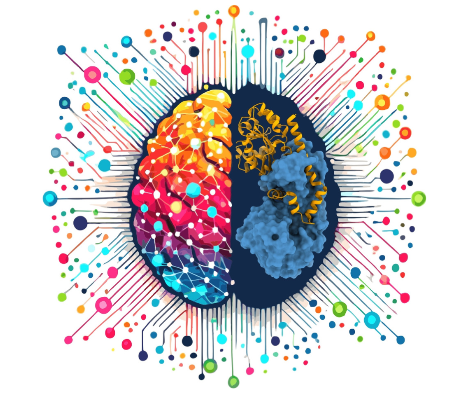
We are made of atoms, just like all living and non-living nature. Atoms combine into molecules. Many of the molecules that function in living organisms are very complex: for example, DNA, RNA, or proteins. To study these molecules at the atomic level, methods are needed that allow us to "magnify" them tens or hundreds of millions of times to visualize their smallest components. This is where the life sciences discipline known as structural biology comes in.
It allows us to understand the chemical reactions underlying the functioning of organisms. We can learn how diseases, including genetic ones, arise or how viruses replicate. Structural biology also enables the visualization of how drugs attach to molecules in a cell. This allows us to optimize this process and consequently create new therapies or improve the properties of existing drugs.
In the Eye of the Synchrotron
The conference was graced by the genius loci of the SOLARIS National Synchrotron Radiation Centre at the Jagiellonian University, the only synchrotron facility in Poland and the first of its kind in this part of Europe. It is the most modern and largest multidisciplinary research device in the country. There are only a few dozen synchrotrons in the world.
These powerful devices, built in a ring shape (Kraków's SOLARIS has a circumference of 96 meters), are electron accelerators and a source of unique light - synchrotron radiation, which allows us to peer into the depths of matter. Attached to the synchrotron are research lines, which can be imagined as extremely efficient microscopes. They allow for the precise capture of any part of reality at the molecular and atomic level. The SOLARIS Centre serves physicists and chemists, and currently, a modern measurement station for protein crystallography is being built on the synchrotron, which will be used by structural biologists. It will serve, among others, in projects aimed at designing new drugs.
"Both structural biology and artificial intelligence technologies are very close to us," said Dr. Jakub Szlachetko, Associate Professor at Jagiellonian University, Director of the SOLARIS Centre. "SOLARIS invests significant resources, including financial, in the development of new research methods that will complement the portfolio of techniques important for structural biology. We are particularly talking about building research lines dedicated to crystallography and X-ray scattering."
Alongside the synchrotron at the SOLARIS Centre operates the Electron Cryomicroscopy Laboratory SOLARIS (Cryo-EM), established thanks to the unprecedented collective effort of the molecular biology community. It has two modern electron cryomicroscopes. The laboratory is one of the best of its kind in Europe, and the research conducted there has become the basis for many publications in the best international scientific journals.
The Electron Breakthrough
For several decades, the dominant method in structural biology was protein crystallography. It involves obtaining microscopic crystals of the studied biological substance, which are then illuminated with X-rays. In the 1990s, the first protein crystallography laboratory was established in Poznań, led by Prof. Mariusz Jaskólski.
These methods did not allow for the understanding of the structure of many important molecules functioning in living organisms, especially those that are very complex and flexible. About ten years ago, there was a significant breakthrough in structural biology, thanks to the development of new methods of electron microscopy that allow the visualization of biological molecules with precision similar to crystallography. In 2017, Jacques Dubochet, Joachim Frank, and Richard Henderson received the Nobel Prize in Chemistry for developing an effective method for generating three-dimensional images of life's molecules. Using electron cryomicroscopy, scientists can now freeze biomolecules in motion and depict them with atomic resolution. However, this method is tedious and time-consuming - determining the structure of a single biological molecule often takes months or years.
The Future Is Now
We are currently experiencing another breakthrough. The Holy Grail of bioinformatics (computational/computer biology) was to develop computational methods for predicting the spatial structures of biological molecules without the need for crystallography or electron microscopy. A milestone towards realizing this dream was reached about two years ago. Current computer predictions of the atomic structure of proteins no longer differ in many cases from structures determined by experimental methods, and their preparation takes seconds, not years. The next challenges are to apply these methods to predict changes in molecule shapes (a feature of many of them) or to identify new substances that could become drugs.
"The dynamic development of structural biology in recent years allows us to understand the molecular basis of diseases," says Miłosz Ruszkowski, Head of the Department of Structural Biology of Eukaryotes at the Institute of Bioorganic Chemistry, Polish Academy of Science. "This makes it possible to develop therapies that are highly effective with minimal side effects. Moreover, we are now on the brink of using structural techniques, such as electron cryotomography, in diagnosing cancers or neurodegenerative diseases. We don't want Poland to miss this opportunity," summarizes the scientist.
The rapid changes occurring in the world of science also create a need for a new approach to educating future biologists. Increasing emphasis will be placed on bioinformatics technologies. In a structural biologist's laboratory, one of the key tools will be a supercomputer with an AI application – thus, increasing investments in IT infrastructure, especially in the context of life sciences, is necessary, the conference participants argued.
"It is clear that the limitations of AI-based techniques mean that they will not replace experimental methods anytime soon. However, they can greatly facilitate and accelerate research in structural biology," said Prof. Marcin Nowotny from IIMCB. "We do not know if computers will provide us with a cure for cancer or an elixir of eternal youth, but certainly new, increasingly effective tools will make it easier, faster, and cheaper to conduct scientific research, which raises hopes for solving the most pressing medical problems in the coming decades," the scientist concluded.
The iNext conference participants agreed that occasional claims that the effects of AI use do not require experimental verification pose a threat to science. As Dr. Piotr Wilk from Jagiellonian University and Claudia Alen Amaro from Instruct-Eric emphasized, funding institutions must be aware that hypotheses generated by AI cannot replace experimental research. They can only accelerate it.

About the iNext Organizers
The meeting in Kraków was organized jointly by European consortia coordinating access to large research infrastructures (scientific equipment platforms): iNext Discovery and INSTRUCT-ERIC. The former includes Polish institutions, while we are striving for access to the latter. The Kraków conference was a milestone in these efforts, being the fruit of combining the forces of both consortia. In one place – the SOLARIS Centre in Kraków – leaders of the most important research infrastructures in Europe, leading structural biologists, and representatives of pioneering companies in the AI revolution in structural biology met.
The iNext-Discovery Foresight Meeting, July 8-10, took place at the SOLARIS National Synchrotron Radiation Centre in Kraków.
International Organizers:
- iNext Discovery
- INSTRUCT
Polish Organizers:
- National Synchrotron Radiation Centre SOLARIS, Jagiellonian University, Kraków
- Institute of Bioorganic Chemistry PAN, Poznań
- International Institute of Molecular and Cell Biology in Warsaw (IIMCB)
- Małopolska Centre of Biotechnology, Kraków
Website: https://inext-foresight.iimcb.gov.pl/
