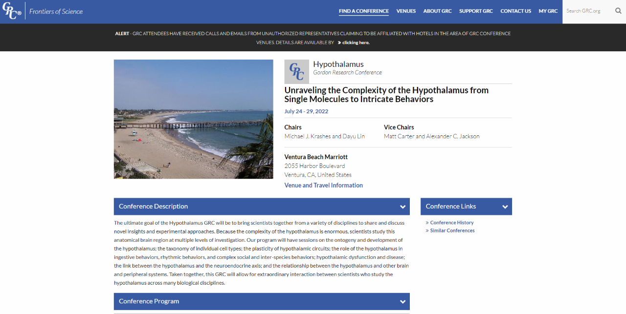
Prof. Andrzej Dziembowski participated in Hypothalamus Gordon Research Conference. Unraveling the Complexity of the Hypothalamus from Single Molecules to Intricate Behaviors; July 24-29, 2022, Ventura, CA, United States with a poster titled "mRNAs of hypothalamic neuropeptades are polyadenylated in the cytoplasm"

thumbnail_image001.png

news---2023-03-08T153340.231-copy.png

We are pleased to inform that in 2022 we established cooperation with the BioCentre of Science Education (BioCEN) in the frame of the GRIEG projects. Our cooperation may cover publicly available laboratory workshops, trainings, presentations and lectures for adolescents, biology teachers, schools, companies and for all who are interested.

BioCentre of Science Education was established in 2002. It is a unique initiative popularizing science. It involves innovative methods, reaches a wide audience, it has its own laboratory in Warsaw and co-organizes events all over Poland for schools, companies, science festivals and other institutions, aiming at reaching teachers and students from smaller towns and villages.

news---2023-03-08T153340.231.png

Miło nam poinformować, że w 2022 roku nawiązaliśmy współpracę z BioCentrum Edukacji Naukowej (BioCEN) w ramach projektów GRIEG. Nasza współpraca może obejmować ogólnodostępne warsztaty laboratoryjne, szkolenia, prezentacje i wykłady dla młodzieży, nauczycieli biologii, szkół, firm i dla wszystkich zainteresowanych.

BioCentrum Edukacji Naukowej powstało w 2002 roku. Jest unikalną inicjatywą popularyzującą naukę. Stosuje innowacyjne metody, dociera do szerokiego grona odbiorców, posiada własne laboratorium w Warszawie oraz współorganizuje wydarzenia w całej Polsce dla szkół, firm, festiwali nauki i innych instytucji, mając na celu dotarcie do nauczycieli i uczniów z mniejszych miast i wsi.


W dniach 18-20 października 2022 roku członkowie Laboratorium Biologii RNA - Grupa ERA Chairs pod kierownictwem prof. Andrzeja Dziembowskiego (koordynatora projektu) spotkali się z partnerami: Centrum Nowych Technologii, Uniwersytet Warszawski (Polska) oraz Uniwersytet w Bergen (Norwegia).

Podczas spotkania omówiono postępy i przyszłe kierunki badań finansowanych z grantu GRIEG. 

IMG 2021

IMG 2022 


On October 18-20, 2022 members of the Laboratory of RNA Biology - ERA Chairs Group headed by Prof. Andrzej Dziembowski (coordinator of the project) met with partners: the Centre of New Technologies, University of Warsaw, Poland and University of Bergen, Norway.

During the meeting progress and future directions of research funded by the GRIEG grant was discussed.


IMG 2021 IMG 2022


Drug development projects

We cooperated with pharmaceutical companies in multiple drug development projects by performing structural studies of complexes of target proteins with inhibitors. Until the end of 2019 these projects were performed by a specialized laboratory – IIMCB Structural Biology Center with Marcin Nowotny as a CSO.

A published example of such cooperation concerns OATD-01 compound developed by OncoArendi Therapeutics company. OATD-01 is an inhibitor chitotriosidase/acidic mammalian chitinase which can be used in the treatment of idiopathic pulmonary fibrosis. The compound recently completed phase I clinical studies. We contributed the structural characterization of the complex between chitinase and the inhibitor.

Robert Koralewski, Barbara Dymek, Marzena Mazur, Piotr Sklepkiewicz, Sylwia Olejniczak, Wojciech Czestkowski, Krzysztof Matyszewski, Gleb Andryianau, Piotr Niedziejko, Michal Kowalski, Mariusz Gruza, Bartłomiej Borek, Karol Jedrzejczak, Agnieszka Bartoszewicz, Elżbieta Pluta, Aleksandra Rymaszewska, Magdalena Kania, Tomasz Rejczak, Sylwia Piasecka, Michal Mlacki, Marcin Mazurkiewicz, Michał Piotrowicz, Magdalena Salamon, Agnieszka Zagozdzon, Agnieszka Napiorkowska-Gromadzka, Aneta Bartlomiejczak, Witold Mozga, Paweł Dobrzański, Karolina Dzwonek, Jakub Golab, Marcin Nowotny, Jacek Olczak, and Adam Golebiowski, Discovery of OATD-01, a First-in-Class Chitinase Inhibitor as Potential New Therapeutics for Idiopathic Pulmonary Fibrosis. J Med Chem 2020 Dec 24;63(24):15527-15540. Published October 20, 2020, https://pubs.acs.org/doi/10.1021/acs.jmedchem.0c01179# 

hCHIT1

Close-up view of active site of hCHIT1 (represented as surface colored according to electrostatic potential) in complex with OATD-01 (blue sticks).


Nucleic acid-based therapeutics

Antisense technology is an emerging therapeutic approach for inhibiting gene expression through recognition and RNase H1-dependent degradation of cellular mRNAs. In this technology, short, synthetic, single-stranded oligonucleotides (ASOs) are designed that are complementary to the target mRNA.

ASOs are frequently chemically modified for enhancing the therapeutic properties of these molecules. The most widely used modifications include phosphorothioate (PS) backbone and modifications of the sugar moieties at 2’ site. The molecular mechanism of toxicity of chemically modified antisense oligonucleotides are not fully understood.

Hyjek-Składanowska M, Vickers T, Napiórkowska A, Anderson BA, Tanowitz M, Crooke ST, Liang X, Seth PP&, Nowotny M&. Origins of the increased affinity of phosphorothioate-modified therapeutic nucleic acids for proteins. J. Am. Chem. Soc., 2020, 142 (16): 7456-7468;
& - corresponding authors

  • The first crystal structure of a complex between a protein and fully PS nucleic acid (DNA-binding domain of a model ASO-binding protein PC4 in complex with full PS 2′-OMe DNA gapmer ASO).

  • The structure reveals a possible mechanism of ASO-induced toxic protein aggregation: ASO is bound in hairpin-like conformation and its exposed gapmer part promotes the formation of a dimer of dimers of PC4 through base pairing.

  • The protein interacts with the PS-nucleic acid through a network of electrostatic and hydrophobic interactions. Importantly, the backbone of the PS ASO is able to form new and more extensive hydrophobic interactions than a natural phosphodiester backbone which provides insights into the origins for the enhanced affinity of PS for proteins.

The studies of PC4-ASO complex have been performed in cooperation with Ionis Pharmaceuticals (Carlsbad, California, USA).

nowotny lab fig13

Structure of PC4 in complex with PS 2′-OMe DNA gapmer ASO. The content of the asymmetric unit of the PTEN ASO complex crystal. PC4 protomers are color-coded and shown in surface representation, DNA is shown in cartoon (PS backbone is shown in yellow, 2′-OMe PS nucleotides are shown in aquamarine, DNA gapmer nucleotides are shown in dark gray). The base pairing between the nucleotides is shown as black dotted lines.


Hyjek-Składanowska M, Anderson BA, Mykhaylyk V, Orr C, Wagner A, Poznański JT, Skowronek K, Seth PP, Nowotny M. Structures of annexin A2-PS DNA complexes show dominance of hydrophobic interactions in phosphorothioate binding. Nucleic Acids Research, 2022;, gkac774, https://doi.org/10.1093/nar/gkac774

  • The first crystal structure of a complex between a PS oligonucleotide and non-specific nucleic acids binding protein (core domain of annexin A2).

  • Interactions between the sulfur atom of the PS linkage and the protein surface are mainly of hydrophobic character - the hydrophobic nature of sulfur contributes to the association of PS ASOs with proteins.

  • Measurements of anomalous diffraction of sulfur confirmed that van der Waals contacts between the sulfur atom and hydrophobic parts of arginine and lysine side chains drive the enhanced interaction of PS ASOs with proteins.

  • Stereoisomer preference at given phosphorothioate in the DNA oligonucleotide is determined by the environment around the PS linkage coming from the protein and other adjacent structural features such as 5-Me groups on cytosine nucleobases.

The studies of AnxA2-ASO complex have been performed in cooperation with Ionis Pharmaceuticals (Carlsbad, California, USA) and Diamond synchrotron (Didcot, Oxfordshire, UK).

nowotny

(A) Crystal structure of AnxA2 in complex with PS ASO duplex. Calcium-binding annexing domain I-IV are coloured light pink, violet, purple, and purple blue, respectively. Calcium ions are shown as orange spheres. DNA is shown in cartoon (PS backbone is shown in yellow, 2’-MOE PS nucleotides are shown in green, DNA gapmer nucleotides are shown in dark gray). (B) Close-up view on the phosphorothioate-binding surface in AnxA2. Polar interactions are shown as red dotted lines. Van der Waals interactions are shown as yellow dotted lines. (C) Difference Fourier anomalous map calculated based on long-wavelength X-ray diffraction data (λ = 2.7552 Ȧ), shown as teal mesh. The preferred occupancy of Rp PS stereoisomer is facilitated by the hydrophobic interactions with surrounding amino acids.


RNA processing

Human cap 2’-OH methyltransferase

mRNA contains a cap structure on its 5' terminus. In higher eukaryotes the first and the second ribonucleotide of the body of the mRNA are methylated on the 2' oxygen. This is thought to serve as a tag to distinguish self mRNA from the mRNA of invading viruses. To circumvent the mechanism, some viruses such as West Nile and yellow fever viruses encode their own 2'-OH methyltransferases.    

Śmietanski M*, Werner M*, Purta E, Kamińska KH, Stepiński J, Darżynkiewicz E, Nowotny M&, Bujnicki JM&. Structural analysis of human 2’-O-ribose methyltransferases involved in mRNA cap structure formation. Nat. Commun., 2014 Jan 9;5:3004; * - equally contributing, & - corresponding authors

  • The first crystal structure of a cellular 2'-OH cap methyltransferase.

  • The mode of cap recognition markedly different from viral counterparts providing hints for antiviral drug development.

The studies of cap 2'-OH methyltransferase have been performed in collaboration with Prof. Janusz M.Bujnicki (IIMCB).

10

Crystal structure of human cap 2'-OH cap methyl transferase 1 (hMTR1). A fragment of capped mRNA is shown in red and the S-adenosylmethionine (methyl group donor) in green.

Mitochondrial exoribonuclease complex mtEXO

RNA degradation pathways play crucial roles in processing of various types of RNA, regulation of gene expression, and efficient removal of defective RNAs. The main executor of RNA turnover and surveillance activity in yeast mitochondria is the mtEXO complex, composed of Dss1 3ʹ-to-5ʹ exoribonuclease and Suv3 helicase.

Rażew M, Warkocki Z, Taube M, Kolondra A, Czarnocki-Cieciura M, Nowak E, Łabędzka-Dmoch K, Kawińska A, Piątkowski J, Golik P, Kozak M, Dziembowski A, Nowotny M. Structural analysis of mtEXO mitochondrial RNA degradosome reveals tight coupling of nuclease and helicase components. Nat Commun., 2018 Jan 8;9(1):97.

  • Crystal structure of Dss1 exoribonuclease from Candida glabrata reveals it is a unique member of the RNase II family with specialized domains responsible for interactions with Suv3 helicase.

  • Crystal structure of the mtEXO complex reveals the arrangement of both subunits in which the helicase motor feeds the 3' end of the RNA into the catalytic channel of Dss1 for its efficient degradation.

  • Co-operation of both helicase and nuclease activities within the complex is particularly important for degeneration of structured RNAs which cannot be handled by Dss1 on its own and for which the unwinding activity of Suv3 is required.


Nowotny Lab Fig. 12

Crystal structure of Candida glabrata mtEXO complex shows the arrangement of the Suv3 helicase on top of Dss1 exoribonucleases and its accessory domains decorating the catalytic RNB domain (shown in blue) with the RNA molecule trapped inside (shown in black).

CutA terminal ribonucleotide transferase

Template-independent terminal ribonucleotide transferases (TENTs) catalyze the addition of nucleotide monophosphates to the 3′-end of RNA molecules regulating their fate. A subgroup TENTs are 3′ CUCU-tagging enzymes, such as CutA in Aspergillus nidulans. CutA preferentially incorporates cytosines, processively polymerizes only adenosines and does not incorporate or extend guanosines.

Malik D, Kobyłecki K, Krawczyk P, Poznański J, Jakielaszek A, Napiórkowska A, Dziembowski A, Tomecki R&, Nowotny M&, Structure and mechanism of CutA, RNA nucleotidyl transferase with an unusual preference for cytosine, Nucleic Acids Res., 2020, 48(16):9387-9405; - corresponding authors

  • The first structural characterization of a TENT adding C/U tails - structures solved for CutA in complex with incoming CTP analog and RNA with three adenosines.

  • The binding of GTP or a primer with a terminal guanosine is predicted to lead to clashes between NH2 of the guanine and the protein, explaining why CutA is unable to use these ligands as substrates.

  • Processive adenosine incorporation likely results from tighter binding of the primer with 3′-terminal adenosine and efficient stacking between adenosine bases of the primer and incoming ATP.

  • A dynamic process of NTP recognition proposed.


nowotny lab 14

Overall structures of CutA complexes. (A) Structure of CutA–CMPCPP complex. The protein is shown as an orange cartoon, with β-strands in a darker shade of orange. Incoming nucleotide is shown as red sticks and active site residues are shown as orange sticks. (B) Structure of CutA–A3 complex. The protein is shown as pink cartoon, with β-strands in purple. The RNA is shown as red sticks and active site residues are shown as pink sticks.

enDR3 – a tool for mapping RNA/DNA hybrids

R-loops are nucleic acid structures consisting of an RNA/DNA hybrid and a displaced single-stranded DNA that arise during transcription. Impaired processing of R-loops has been linked to genome instability, a hallmark of various severe human diseases. Despite their biological importance, the mechanisms governing R-loop formation and regulation remain poorly understood and new tools are needed to their precise localization in the genome.

Marta Jedynak-Slyvka*, Zuzanna Kaczmarska*, Damian Graczyk, Aneta Jurkiewicz, Małgorzata Figiel, Lukasz S Borowski, Roman J Szczesny&, Marcin Nowotny& Genome-wide in vivo and ex vivo mapping of R-loops using engineered N-terminal hybrid-binding domain of RNase H3 (enDR3), Nucleic Acids Res., 2025: 53(15); * - equally contributing, & - corresponding authors

  • enDR3 is a novel tool engineered to capture and sequence RNA/DNA hybrids with high precision for accurate R-loop localization.

  • It consists of an engineered tandem fusion of a modified N-terminal hybrid-binding domain derived from bacterial RNase H3.

  • enDR3 has been successfully utilized for genome-wide R-loop profiling through DNA/RNA immunoprecipitation coupled with complementary DNA conversion and chromatin immunoprecipitation techniques.

  • The tool enables robust detection of R-loops both ex vivo and in vivo, offering a versatile platform for advancing research in R-loop biology.

abstract1

Schematic of the use of enDR3 in mapping RNA/DNA hybrids (R-loops) ex vivo and in vivo


Reverse transcriptases

XMRV reverse transcriptase

Reverse transcriptases use two enzymatic activities – DNA polymerase and RNase H to catalyze the conversion of single-stranded RNA to double-stranded DNA, a process essential for proliferation of retroviruses such as HIV and retrotransposons. Retroviral RTs are divided into two classes – dimeric (i. e. HIV) or monomeric (i. e. gammaretroviral enzyme from mouse Moloney leukemia virus and closely related XMRV).

Nowak E, Potrzebowski W, Konarev PV, Rausch JW, Bona MK, Svergun DI, Bujnicki JM, Le Grice SF, Nowotny M. Structural analysis of monomeric retroviral reverse transcriptase in complex with an RNA/DNA hybrid. Nucleic Acids Res., 2013 Apr 1;41(6):3874-87

  • The first crystal structure of a monomeric RTs in complex with RNA/DNA hybrid visualizing the polymerase-connection fragment of the enzyme.

  • Full-length protein modelled based on small-angle X-ray scattering data.

  • SAXS data demonstrated that the RNase H domain is mobile and only occasionally interacts with the substrate to cleave RNA. This is the mechanism to fine tune RNase H activity.

  • This mechanism of RNase H fine-tuning is different from dimeric RTs which use substrate deformations for that purpose.

8

Model of the full length XMRV RT based on SAXS data, a crystal structure of the polymerase-connection fragment in complex with RNA/DNA hybrid (blue-fingers, red-palm, green-thumb, yellow-connection) and the structure of isolated XMRV RNase H domain (orange, Zhou D, J Struct Biol. 2012). RNA template stand is in purple and DNA primer strand in blue.

Foamy viral protease-reverse transcriptase

One of the distinguishing features of foamy viruses (FV) is the different way of replication. Additionally they synthesize a separate mRNA from which the Pol protein is expressed as a precursor of PR-RT-IN, which is later cleaved into two proteins: PR-RT and IN. Thus, FV RT is the only protein of its type to contain an additional PR domain located at the N-terminus of the protein.

Nowacka M*, Nowak E*&, Czarnocki-Cieciura M, Jackiewicz J, Skowronek K, Szczepanowski RH, Wöhrl BM, Nowotny M&. Structures of substrate complexes of foamy viral protease-reverse transcriptase. J Virol., 2021; 95(18): e0084821. *- equally contributing, & - corresponding authors

  • First structures of substrate complexes of a foamy viral protease-reverse transcriptase. 

  • Novel mechanism of switching between protein monomer on RNA/DNA substrate and dimer on dsDNA.  

  • Mobile RNase H domain of foamy viral PR-RT transiently interacts with the RNA/DNA substrate 18-21 nt from the polymerase active site.


overall fig annual3 website

Structures of nucleic acid complexes of MFV PR-RT. (A) Crystal structure of MFV PR-RT in complex with an RNA/DNA hybrid substrate. (B) Cryo-EM structure of MFV PR-RT ΔRH in complex with dsDNA. Lighter shades of colors are used for domains/subdomains of subunit B.


Ty3 reverse transcriptase

Retrotransposons are mobile genetic elements that replicate with an RNA intermediate. Reverse activity of element-encoded RT is essential for this process. Retroelements are one of the most potent forces shaping eukaryotic genomes – more than 40% of human genome derives from those elements. Ty3 is a yeast retrolement from long-terminal class termed Ty3/Copia. It is thought that retroviruses evolved from this class of retrotransposons.


Nowak E, Miller JT, Bona MK, Studnicka J, Szczepanowski RH, Jurkowski J, Le Grice SFJ&, Nowotny M&. Ty3 reverse transcriptase complexed with an RNA-DNA hybrid shows structural and functional asymmetry. Nature Struct. Mol. Biol., 2014; 21(4):389-96;
& - corresponding authors

  • The first crystal structure of a retrotransposon RT.

  • Ty3 RT forms an asymmetric homodimer in which one subunit has the polymerase competent configuration and the other has an altered conformation and harbors the RNase H activity.

  • RNase H is postulated to undergo a conformational change to reach the position required for RNA/DNA cleavage, which regulates this activity.

  • Ty3 and HIV RT architecture differs: HIV enzyme is a constitutive heterodimer with both polymerase and RNase H activities residing in the larger subunit and Ty3 is a substrate-induced homodimer in which the two activities are located in different subunits.

  • The studies of XMRV and Ty3 reverse transcriptases have been performed in collaboration with Dr. Stuart Le Grice (National Institutes of Health, USA).


images/labs/nowotny/9.jpg
Crystal structure of Ty3 reverse transcriptase. The subunit with polymerase-competent configuration is shown in darker color (blue-fingers, red-palm, green-thumb, yellow-RNase H) and the subunit with altered conformation in lighter shades of the same colors. RNA template stand is in purple and DNA primer strand in blue. Active site residues for polymerase and RNase H domain are shown as sticks.


Cauliflower mosaic virus reverse transcriptase (CaMV RT)

CaMV is a prominent plant virus, important in both plant pathology and agricultural biotechnology. Even though the genome of CaMV is a double-stranded DNA, it replicates through reverse transcription. Interestingly, the structural features of CaMV RT make it similar to Ty3 RT, but its mode of action and enzymatic properties differ from other reverse transcriptases.

Chandrasekaran Prabaharan, Małgorzata Figiel, Roman H. Szczepanowski, Krzysztof Skowronek, Weronika Zajko, Vinuchakkaravarthy Thangaraj, Sebastian Chamera, Elzbieta Nowak, and Marcin Nowotny, Structural and biochemical characterization of cauliflower mosaic virus reverse transcriptase, J. Biol. Chem. (2024) 300(8), 107555.

  • The first crystal structure of a plant viral RT in complex with an RNA/DNA hybrid substrate.

  • CaMV RT forms a monomeric complex with the hybrid substrate and adopts a polymerase configuration. A single molecule of CaMV RT performs DNA synthesis.

  • Novel mechanism of RNase H activity that requires two copies of the enzyme for RNA hydrolysis. One molecule is involved in structural stabilisation and another forms a transient association with the monomeric complex for RNA cleavage.

CaMV
Crystal structure ofCaMV RT-RNA/DNA hybrid complex. Domains of CaMV RT are labelled and RNA and DNA strands are shown in magenta and cyan, respectivel


HIV-1 reverse transriptase

HIV-1 RT is an important drug target in therapy of HIV-1 infection. The RT uses its polymerase and RNase H activities to catalyze the process of reverse transcription, in which the single-stranded RNA of the virus is converted into double-stranded DNA that can be integrated into the host cell genome.

Figiel M, Krepl M, Poznański J, Gołąb A, Šponer J, Nowotny M. Coordination between the polymerase and RNase H activity of HIV-1 reverse transcriptase. Nucleic Acids Res., 2017; 45(6):3341-3352.

  • Dynamics of HIV-1 RT-RNA/DNA complex were studied using a combination of chemical cross-linking and molecular dynamics simulations.

  • The RNA/DNA substrate can simultaneously interact with the polymerase and RNase H active sites.

  • Untwisting of the RNA/DNA substrate double helix is required for its productive interaction with the RNase H active site.

  • This allows HIV-1 RT to regulate the amount of the RNase H activity.


hiv1 nowotny lab

Molecular dynamics simulation of HIV-1 RT RNA/DNA complex (two views). Superposition of the starting model (light colors) and the final model in MD simulations (dark colors). Domains of HIV-1 RT are labeled. RNA and DNA strands of the substrate are shown in red and blue, respectively.


In order to be integrated into the host cell genome, the single-stranded RNA of HIV-1 is converted into double-stranded DNA. The synthesis of the (+)-strand DNA starts from the polypurine tract (PPT) primer. The PPT primer is generated by the RNase H domain of HIV-1 RT which cuts specifically at its termini, but leaves the body of the PPT intact.

Figiel M, Krepl M, Park S, Poznański J, Skowronek K, Gołąb A, Ha T, Šponer J, Nowotny M. Mechanism of polypurine tract primer generation by HIV-1 reverse transcriptase. J Biol. Chem., 2018; 293(1):191-202.

  • Factors involved in recognition of the PPT sequence by HIV-1 RT-RNA/DNA were studied using a combination of chemical cross-linking, molecular dynamics simulations, and single-molecule assays.

  • The PPT is specifically recognized after the complex with HIV-1 RT is formed and not at the stage of binding.

  • Recognition of the PPT is based on two elements: agreement with the sequence preference of RNase H and the indirect readout of the poly-rA/dT stretch. The rigid but brittle poly-rA/dT is not compatible with the catalytically relevant substrate geometry and is prone to undergo sequence slippage upon deformation.

  • The poor match with the RNase H sequence preference and the dynamic properties of the PPT explain the protection of its body from cleavage.

Studies of HIV-1 RT have been done in collaboration with Jiří Šponer (Institute of Biophysics, CAS) and Taekjip Ha (Johns Hopkins Univ.).


Nowotny Fig. 11

Model of PPT recognition by HIV-1 RT. Cleavage at the expected site involves both preferred residues at the cleavage consensus position and the ability of PPT sequence to undergo the appropriate conformational change without distortion (left panel). Cleavage in the middle of the PPT body (A-tract) is inhibited by three elements: non-preferred residues in the consensus positions, rigidity of the poly(rA-dT) sequence, and misalignment of the substrate at the RNAse H active site due to poly(rA-dT) sequence slippage

Abi polymerases 

Abortive infection (Abi) is a bacterial antiphage defense strategy involving suicide of the infected cell. Some Abi pathways involve polymerases that are related to reverse transcriptases and combine the ability to synthesize DNA in a template-independent manner with protein priming. The first reverse transcriptase-related proteins that were proposed to utilize the abortive infection antiphage strategy were AbiK, Abi-P2 and AbiA. Within this set, AbiA is the only protein to contain an additional component – a higher eukaryotes and prokaryotes nucleotide-binding (HEPN) domain which is also found in several other systems of antiviral defense.

Figiel M*&, Gapińska M&, Czarnocki-Cieciura M, Zajko W, Sroka M, Skowronek K, Nowotny M*. Mechanism of protein-primed template-independent DNA synthesis by Abi polymerases. Nucleic Acids Res., 2022; 50(17):10026-10040. 

& - equally contributing; * - corresponding authors 

  • Cryo-EM and crystal structure of AbiK-DNA adduct, cryo-EM structure of protein-priming deficient AbiK variant, crystal structure of Abi-P2 

  • AbiK-DNA structure is the first one showing a protein-DNA adduct resulting from protein-primed DNA synthesis 

  • AbiK and Abi-P2 are the first examples of RT-related enzymes that adopt hexameric or trimeric architecture; the oligomerization is required for enzymatic activity 

  • AbiK and Abi-P2 adopt a bilobal structure with an RT-like domain that comprises palm and fingers subdomains and a unique helical domain 

  • Protein priming by Ll-AbiK involves a conformational change of a mobile loop harboring the priming tyrosine residue 

Abi crystal

Crystal structures of AbiK-DNA adduct (left) and Abi-P2 (right) polymerases shown in surface representation. Individual subunits are shown in different colors, DNA strands are shown as black cartoon. 

Gapińska M, Zajko W, Skowronek K, Figiel M, Krawczyk PS, Egorov AA, Dziembowski A, Johansson MJO*, Nowotny M*. Structure-functional characterization of Lactococcus AbiA phage defense system. Nucleic Acids Res., 2024, 52(8):4723-4738.
* - corresponding authors

  • a crystal structure of AbiA-DNA complex

  • AbiA forms dimers and HEPN domain is essential for dimerization

  • AbiA exhibits template-independent DNA polymerase activity with nucleotide preference for adenosines and cytosines

  • HEPN domain is enzymatically inactive, yet its presence is required for DNA polymerase activity of AbiAProtein priming by Ll-AbiK involves a conformational change of a mobile loop harboring the priming tyrosine residue 

  • DNA polymerase activity of AbiA is essential for protection against invading phages as evidenced by an infection assay in E. coli surrogate host

  • contrary to previous assumptions, the antiphage defense mediated by AbiA and AbiK appears not to rely on induced cell death

Studies of AbiA were performed in collaboration with Marcus Johansson (Lund University, Sweden) and Andrzej Dziembowski (IIMCB).

AbiaA(left) Crystal structure of L. lactis AbiA protein dimer. Domains of one of the subunits are colored and labeled, DNA product of the enzyme is shown in green.
(right) Results of the infection assay, in which E. coli cells harboring wild-type and mutated versions of AbiA were challenged by different phages. Formation of plaques indicates successful infection.