
Reverse transcriptases
XMRV reverse transcriptase
Reverse transcriptases use two enzymatic activities – DNA polymerase and RNase H to catalyze the conversion of single-stranded RNA to double-stranded DNA, a process essential for proliferation of retroviruses such as HIV and retrotransposons. Retroviral RTs are divided into two classes – dimeric (i. e. HIV) or monomeric (i. e. gammaretroviral enzyme from mouse Moloney leukemia virus and closely related XMRV).
Nowak E, Potrzebowski W, Konarev PV, Rausch JW, Bona MK, Svergun DI, Bujnicki JM, Le Grice SF, Nowotny M. Structural analysis of monomeric retroviral reverse transcriptase in complex with an RNA/DNA hybrid. Nucleic Acids Res., 2013 Apr 1;41(6):3874-87
-
The first crystal structure of a monomeric RTs in complex with RNA/DNA hybrid visualizing the polymerase-connection fragment of the enzyme.
-
Full-length protein modelled based on small-angle X-ray scattering data.
-
SAXS data demonstrated that the RNase H domain is mobile and only occasionally interacts with the substrate to cleave RNA. This is the mechanism to fine tune RNase H activity.
-
This mechanism of RNase H fine-tuning is different from dimeric RTs which use substrate deformations for that purpose.
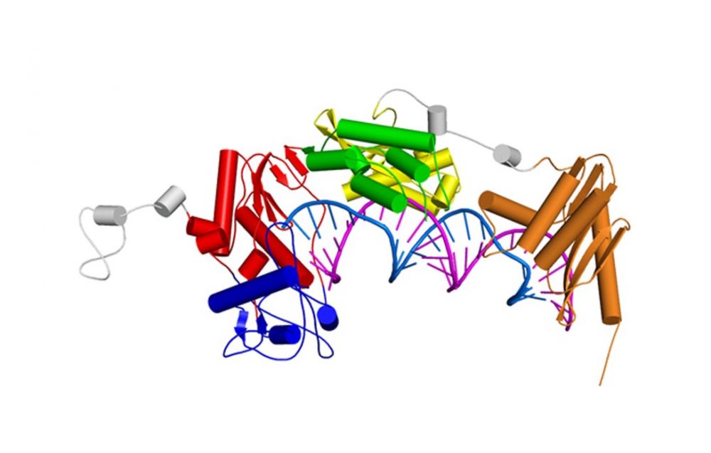
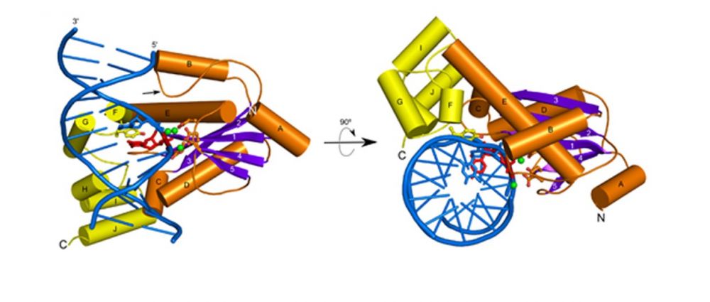
Model of the full length XMRV RT based on SAXS data, a crystal structure of the polymerase-connection fragment in complex with RNA/DNA hybrid (blue-fingers, red-palm, green-thumb, yellow-connection) and the structure of isolated XMRV RNase H domain (orange, Zhou D, J Struct Biol. 2012). RNA template stand is in purple and DNA primer strand in blue.
Foamy viral protease-reverse transcriptase
One of the distinguishing features of foamy viruses (FV) is the different way of replication. Additionally they synthesize a separate mRNA from which the Pol protein is expressed as a precursor of PR-RT-IN, which is later cleaved into two proteins: PR-RT and IN. Thus, FV RT is the only protein of its type to contain an additional PR domain located at the N-terminus of the protein.
Nowacka M*, Nowak E*&, Czarnocki-Cieciura M, Jackiewicz J, Skowronek K, Szczepanowski RH, Wöhrl BM, Nowotny M&. Structures of substrate complexes of foamy viral protease-reverse transcriptase. J Virol., 2021; 95(18): e0084821. *- equally contributing, & - corresponding authors
-
First structures of substrate complexes of a foamy viral protease-reverse transcriptase.
-
Novel mechanism of switching between protein monomer on RNA/DNA substrate and dimer on dsDNA.
-
Mobile RNase H domain of foamy viral PR-RT transiently interacts with the RNA/DNA substrate 18-21 nt from the polymerase active site.

Structures of nucleic acid complexes of MFV PR-RT. (A) Crystal structure of MFV PR-RT in complex with an RNA/DNA hybrid substrate. (B) Cryo-EM structure of MFV PR-RT ΔRH in complex with dsDNA. Lighter shades of colors are used for domains/subdomains of subunit B.
Ty3 reverse transcriptase
Retrotransposons are mobile genetic elements that replicate with an RNA intermediate. Reverse activity of element-encoded RT is essential for this process. Retroelements are one of the most potent forces shaping eukaryotic genomes – more than 40% of human genome derives from those elements. Ty3 is a yeast retrolement from long-terminal class termed Ty3/Copia. It is thought that retroviruses evolved from this class of retrotransposons.
Nowak E, Miller JT, Bona MK, Studnicka J, Szczepanowski RH, Jurkowski J, Le Grice SFJ&, Nowotny M&. Ty3 reverse transcriptase complexed with an RNA-DNA hybrid shows structural and functional asymmetry. Nature Struct. Mol. Biol., 2014; 21(4):389-96;
& - corresponding authors
-
The first crystal structure of a retrotransposon RT.
-
Ty3 RT forms an asymmetric homodimer in which one subunit has the polymerase competent configuration and the other has an altered conformation and harbors the RNase H activity.
-
RNase H is postulated to undergo a conformational change to reach the position required for RNA/DNA cleavage, which regulates this activity.
-
Ty3 and HIV RT architecture differs: HIV enzyme is a constitutive heterodimer with both polymerase and RNase H activities residing in the larger subunit and Ty3 is a substrate-induced homodimer in which the two activities are located in different subunits.
-
The studies of XMRV and Ty3 reverse transcriptases have been performed in collaboration with Dr. Stuart Le Grice (National Institutes of Health, USA).

Crystal structure of Ty3 reverse transcriptase. The subunit with polymerase-competent configuration is shown in darker color (blue-fingers, red-palm, green-thumb, yellow-RNase H) and the subunit with altered conformation in lighter shades of the same colors. RNA template stand is in purple and DNA primer strand in blue. Active site residues for polymerase and RNase H domain are shown as sticks.
Cauliflower mosaic virus reverse transcriptase (CaMV RT)
CaMV is a prominent plant virus, important in both plant pathology and agricultural biotechnology. Even though the genome of CaMV is a double-stranded DNA, it replicates through reverse transcription. Interestingly, the structural features of CaMV RT make it similar to Ty3 RT, but its mode of action and enzymatic properties differ from other reverse transcriptases.
Chandrasekaran Prabaharan, Małgorzata Figiel, Roman H. Szczepanowski, Krzysztof Skowronek, Weronika Zajko, Vinuchakkaravarthy Thangaraj, Sebastian Chamera, Elzbieta Nowak, and Marcin Nowotny, Structural and biochemical characterization of cauliflower mosaic virus reverse transcriptase, J. Biol. Chem. (2024) 300(8), 107555.
-
The first crystal structure of a plant viral RT in complex with an RNA/DNA hybrid substrate.
-
CaMV RT forms a monomeric complex with the hybrid substrate and adopts a polymerase configuration. A single molecule of CaMV RT performs DNA synthesis.
-
Novel mechanism of RNase H activity that requires two copies of the enzyme for RNA hydrolysis. One molecule is involved in structural stabilisation and another forms a transient association with the monomeric complex for RNA cleavage.
Crystal structure ofCaMV RT-RNA/DNA hybrid complex. Domains of CaMV RT are labelled and RNA and DNA strands are shown in magenta and cyan, respectivel
HIV-1 reverse transriptase
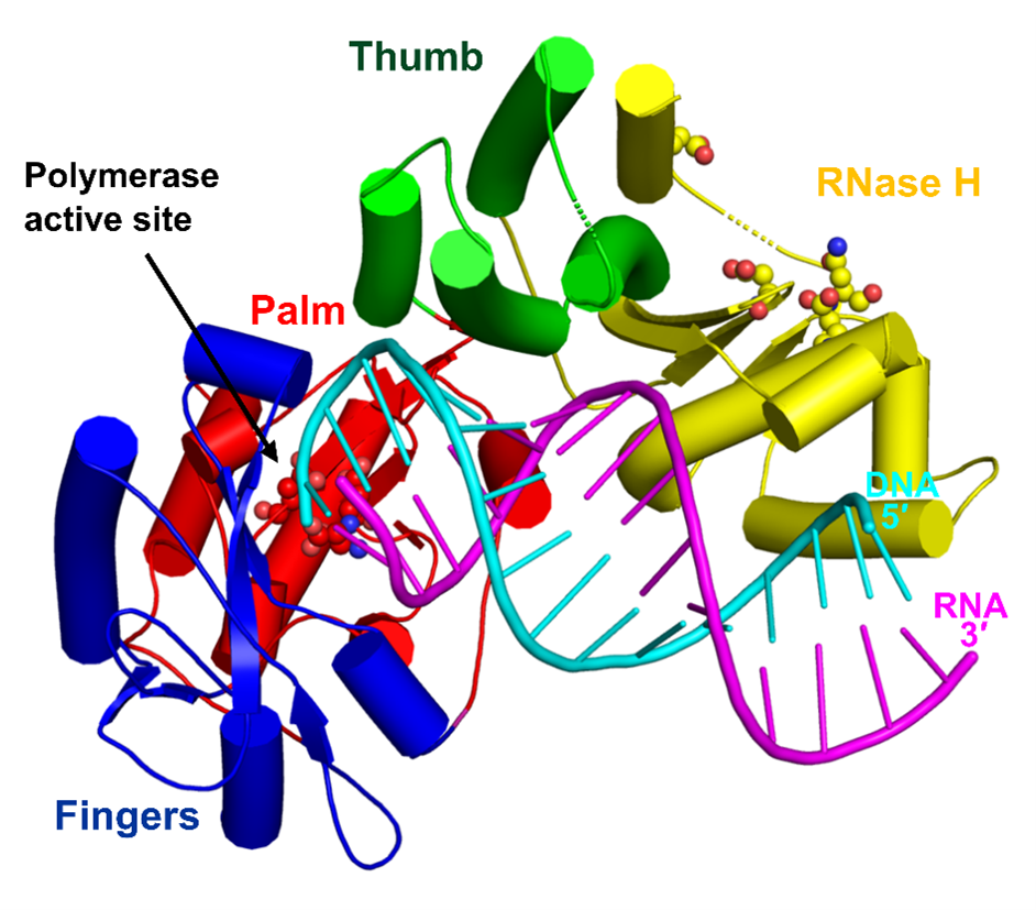
HIV-1 RT is an important drug target in therapy of HIV-1 infection. The RT uses its polymerase and RNase H activities to catalyze the process of reverse transcription, in which the single-stranded RNA of the virus is converted into double-stranded DNA that can be integrated into the host cell genome.
Figiel M, Krepl M, Poznański J, Gołąb A, Šponer J, Nowotny M. Coordination between the polymerase and RNase H activity of HIV-1 reverse transcriptase. Nucleic Acids Res., 2017; 45(6):3341-3352.
-
Dynamics of HIV-1 RT-RNA/DNA complex were studied using a combination of chemical cross-linking and molecular dynamics simulations.
-
The RNA/DNA substrate can simultaneously interact with the polymerase and RNase H active sites.
-
Untwisting of the RNA/DNA substrate double helix is required for its productive interaction with the RNase H active site.
-
This allows HIV-1 RT to regulate the amount of the RNase H activity.
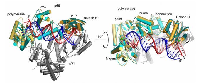
Molecular dynamics simulation of HIV-1 RT RNA/DNA complex (two views). Superposition of the starting model (light colors) and the final model in MD simulations (dark colors). Domains of HIV-1 RT are labeled. RNA and DNA strands of the substrate are shown in red and blue, respectively.
In order to be integrated into the host cell genome, the single-stranded RNA of HIV-1 is converted into double-stranded DNA. The synthesis of the (+)-strand DNA starts from the polypurine tract (PPT) primer. The PPT primer is generated by the RNase H domain of HIV-1 RT which cuts specifically at its termini, but leaves the body of the PPT intact.
Figiel M, Krepl M, Park S, Poznański J, Skowronek K, Gołąb A, Ha T, Šponer J, Nowotny M. Mechanism of polypurine tract primer generation by HIV-1 reverse transcriptase. J Biol. Chem., 2018; 293(1):191-202.
-
Factors involved in recognition of the PPT sequence by HIV-1 RT-RNA/DNA were studied using a combination of chemical cross-linking, molecular dynamics simulations, and single-molecule assays.
-
The PPT is specifically recognized after the complex with HIV-1 RT is formed and not at the stage of binding.
-
Recognition of the PPT is based on two elements: agreement with the sequence preference of RNase H and the indirect readout of the poly-rA/dT stretch. The rigid but brittle poly-rA/dT is not compatible with the catalytically relevant substrate geometry and is prone to undergo sequence slippage upon deformation.
-
The poor match with the RNase H sequence preference and the dynamic properties of the PPT explain the protection of its body from cleavage.
Studies of HIV-1 RT have been done in collaboration with Jiří Šponer (Institute of Biophysics, CAS) and Taekjip Ha (Johns Hopkins Univ.).
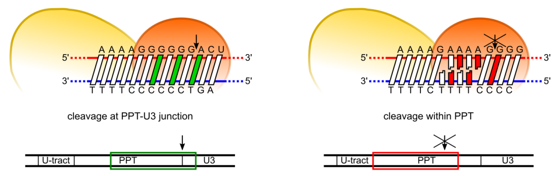
Model of PPT recognition by HIV-1 RT. Cleavage at the expected site involves both preferred residues at the cleavage consensus position and the ability of PPT sequence to undergo the appropriate conformational change without distortion (left panel). Cleavage in the middle of the PPT body (A-tract) is inhibited by three elements: non-preferred residues in the consensus positions, rigidity of the poly(rA-dT) sequence, and misalignment of the substrate at the RNAse H active site due to poly(rA-dT) sequence slippage
Abi polymerases
Abortive infection (Abi) is a bacterial antiphage defense strategy involving suicide of the infected cell. Some Abi pathways involve polymerases that are related to reverse transcriptases and combine the ability to synthesize DNA in a template-independent manner with protein priming. The first reverse transcriptase-related proteins that were proposed to utilize the abortive infection antiphage strategy were AbiK, Abi-P2 and AbiA. Within this set, AbiA is the only protein to contain an additional component – a higher eukaryotes and prokaryotes nucleotide-binding (HEPN) domain which is also found in several other systems of antiviral defense.
Figiel M*&, Gapińska M&, Czarnocki-Cieciura M, Zajko W, Sroka M, Skowronek K, Nowotny M*. Mechanism of protein-primed template-independent DNA synthesis by Abi polymerases. Nucleic Acids Res., 2022; 50(17):10026-10040.
& - equally contributing; * - corresponding authors
-
Cryo-EM and crystal structure of AbiK-DNA adduct, cryo-EM structure of protein-priming deficient AbiK variant, crystal structure of Abi-P2
-
AbiK-DNA structure is the first one showing a protein-DNA adduct resulting from protein-primed DNA synthesis
-
AbiK and Abi-P2 are the first examples of RT-related enzymes that adopt hexameric or trimeric architecture; the oligomerization is required for enzymatic activity
-
AbiK and Abi-P2 adopt a bilobal structure with an RT-like domain that comprises palm and fingers subdomains and a unique helical domain
-
Protein priming by Ll-AbiK involves a conformational change of a mobile loop harboring the priming tyrosine residue
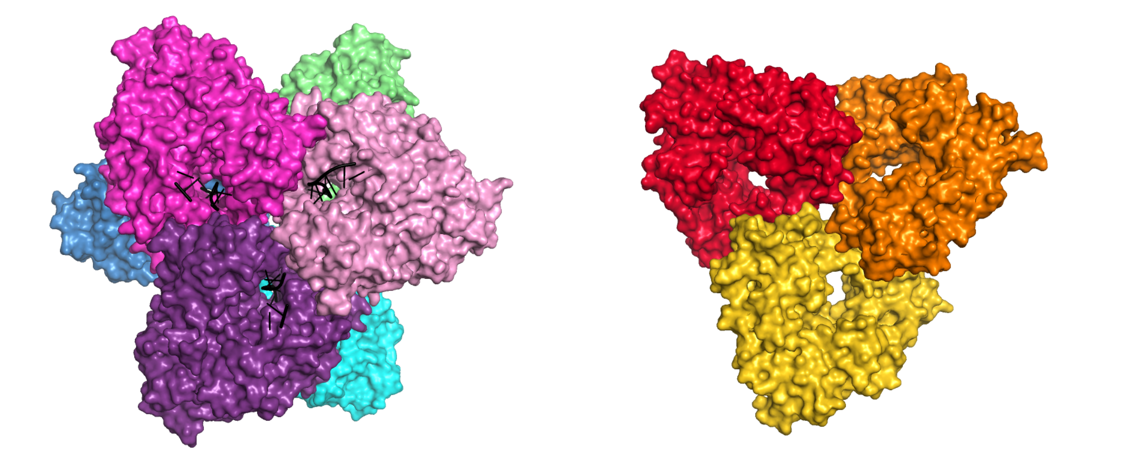
Crystal structures of AbiK-DNA adduct (left) and Abi-P2 (right) polymerases shown in surface representation. Individual subunits are shown in different colors, DNA strands are shown as black cartoon.
Gapińska M, Zajko W, Skowronek K, Figiel M, Krawczyk PS, Egorov AA, Dziembowski A, Johansson MJO*, Nowotny M*. Structure-functional characterization of Lactococcus AbiA phage defense system. Nucleic Acids Res., 2024, 52(8):4723-4738.
* - corresponding authors
-
a crystal structure of AbiA-DNA complex
-
AbiA forms dimers and HEPN domain is essential for dimerization
-
AbiA exhibits template-independent DNA polymerase activity with nucleotide preference for adenosines and cytosines
-
HEPN domain is enzymatically inactive, yet its presence is required for DNA polymerase activity of AbiAProtein priming by Ll-AbiK involves a conformational change of a mobile loop harboring the priming tyrosine residue
-
DNA polymerase activity of AbiA is essential for protection against invading phages as evidenced by an infection assay in E. coli surrogate host
-
contrary to previous assumptions, the antiphage defense mediated by AbiA and AbiK appears not to rely on induced cell death
Studies of AbiA were performed in collaboration with Marcus Johansson (Lund University, Sweden) and Andrzej Dziembowski (IIMCB).
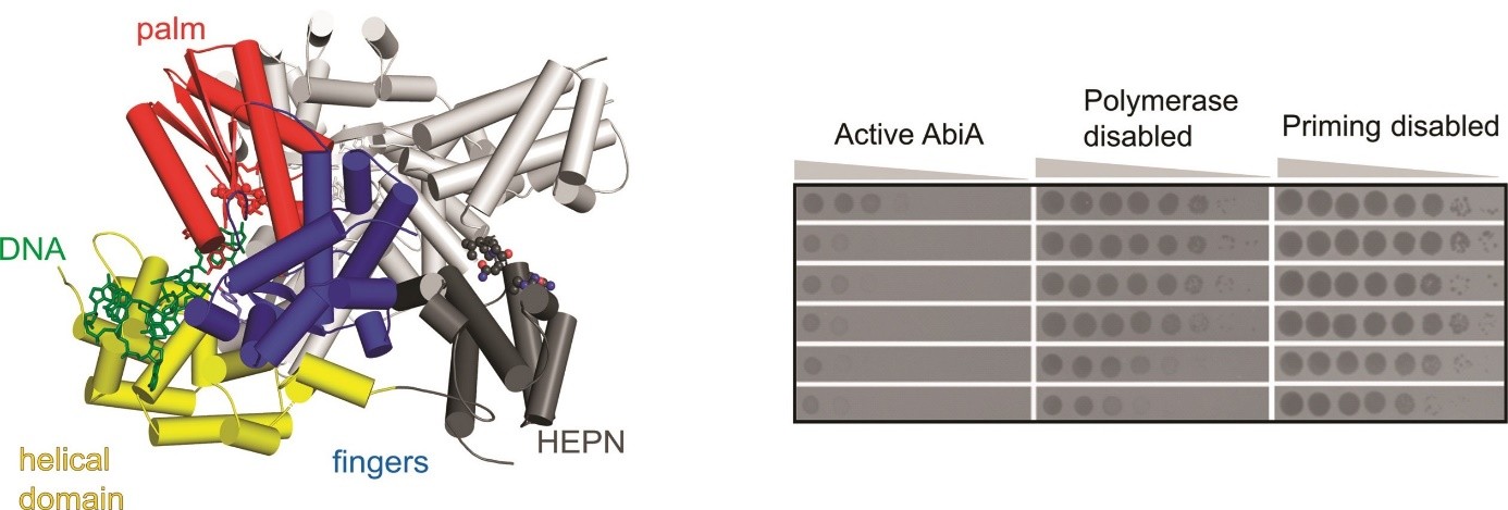 (left) Crystal structure of L. lactis AbiA protein dimer. Domains of one of the subunits are colored and labeled, DNA product of the enzyme is shown in green.
(right) Results of the infection assay, in which E. coli cells harboring wild-type and mutated versions of AbiA were challenged by different phages. Formation of plaques indicates successful infection.
DNA transposition
Tn7 Transposase TnsB
Bacterial Tn7 elements are among the best-studied and most widespread DNA transposons and are detected in 10%–20% of sequenced bacterial genomes. Tn7 mobility is tightly controlled and mediated by five element-encoded proteins. Translocation of the Tn7 is executed by a heteromeric transposase TnsA+TnsB. The TnsB transposase recognizes the left and right ends of the element, catalyzes cleavage at the 3′ ends of the transposon, and performs the strand-transfer reaction that leads to the insertion of the element into a target DNA.
Kaczmarska Z*, Czarnocki-Cieciura M*, Górecka-Minakowska KM*, Wingo RJ, Jackiewicz J, Zajko W, Poznański JT, Rawski M, Grant T, Peters JE&, Nowotny M.& Structural basis of transposon end recognition explains central features of Tn7 transposition systems. Mol Cell, 2022 Jul 21;82(14):2618-2632.e7
* - equally contributing, & - corresponding authors
-
Cryo-EM structure of Tn7 transposase TnsB in complex with transposon end DNA
-
TnsB chains interact with its binding sites in DNA in a tiled and intertwined fashion
-
TnsB exhibits a DNA sequence preference rather than strict specificity
-
Overlap of TnsB-binding sites in the right Tn7 end is key for its functionality
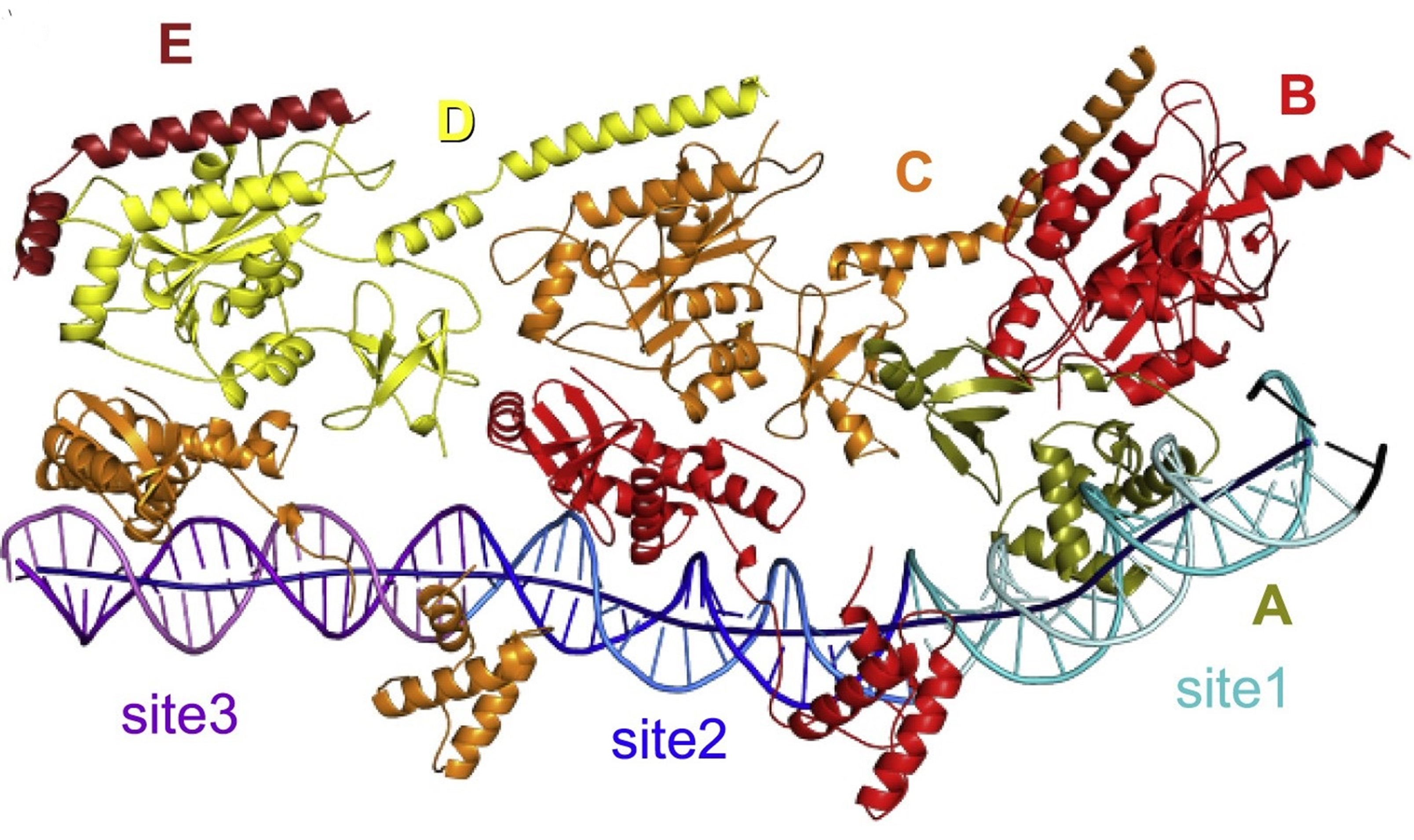
Beads-on-a-string organization of TnsB bound to the fragment of the right transposon end. The TnsB chains (labeled with capital letters) and DNA sites are colored as follows: olive for chain A, red for chain B, orange for chain C, yellow for chain D, burgundy for chain E, light cyan/cyan (top/bottom strands) for site 1, blue/dark blue for site 2, pink/purple for site 3, and black for transposon terminus. The axis of the DNA helix is shown as a dark blue tube.
DNA repair
Bacterial Nucleotide Excision Repair
Nucleotide excision repair (NER) is one of the major pathways of DNA repair and its main feature is its ability to recognize a wide spectrum of DNA lesions of various sizes and structures. In bacterial NER UvrA, a dimeric ATPase, plays the role of a DNA damage sensor. Damage verification is performed by UvrB helicase and UvrC double nuclease excises the damaged DNA fragment.
Jaciuk M, Nowak E, Skowronek K, Tanska A, Nowotny M. Structure of UvrA nucleotide excision repair protein in complex with modified DNA. Nature Struct. Mol. Biol., 2011; 18:191-197
-
The first crystal structure of UvrA interacting with damaged dsDNA obtained using the protein from T. maritima.
-
UvrA does not interact with the damage site directly but senses the deformed conformation of the DNA induced by the presence of the lesion – bending, stretching and unwinding.
Crystal structure of UvrA dimer (one monomer in color, the other in gray). One ATPase module is shown in cyan and red and the other in pink and blue. The DNA-binding domain is in green and UvrB-binding domain in yellow. Structural zinc ions are shown as orange spheres.
Jaciuk M*, Swuec P*, Gaur V*, Kasprzak JM, Renault L, Dobrychłop M, Nirwal S, Bujnicki JM& , Costa A&, Nowotny M&. A combined structural and biochemical approach reveals translocation and stalling of UvrB on the DNA lesion as a mechanism of damage verification in bacterial nucleotide excision repair. DNA Repair, 2019; 85, 102746; * - equally contributing, & - corresponding authors
-
Computational structural model of UvrA—UvrB—DNA complex involved in DNA damage verification in bacterial DNA repair was prepared and corroborated experimentally by electron microscopy.
-
UvrB uses a β-hairpin element to clamp one DNA strand, each one of the two UvrB molecules in the complex clamps a different DNA strand.
-
UvrB translocates in 3′ direction toward the DNA lesion where the UvrB molecule clamping the damaged strand stalls and recruits UvrC nuclease.
-
This mechanism explains how the initial imprecise localization of the damage by UvrA is converted to precise and strand-specific localization to promote accurate incisions by UvrC.
The studies of damage verification in bacterial NER have been performed in collaboration with Janusz M. Bujnicki (IIMCB) and Alessandro Costa (The Crick Institute).
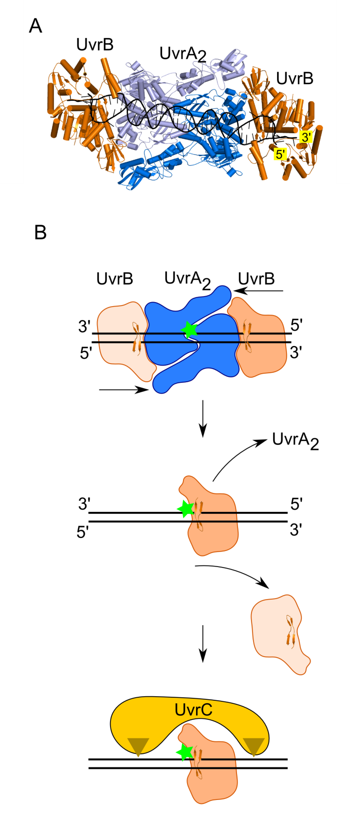
Model of damage verification in bacterial NER. (A) Model of UvrA2—UvrB2—DNA complex. UvrA dimer shown in two shades of blue. UvrB shown in orange. DNA shown in black. (B) Proposed mechanism. The UvrA dimer bound at the site of DNA modification recruits two UvrB molecules. Each UvrB molecule clamps a different DNA strand under the B-hairpin element (upper panel). Both UvrB molecules then translocate toward the lesion with 5′ to 3′ polarity on the strand under the hairpin. The UvrB molecule that clamps the modified strand will stall at the lesion (green star indicates the site of DNA modification) and the other UvrB molecule (light orange) will dissociate (middle panel). The stalled UvrB recruits UvrC double nuclease (shown in yellow), which makes two incisions indicated with triangles.
Rad2
Eukaryotic nucleotide excision repair (NER) is one of the major DNA repair pathways. It involves the excision of the DNA fragment containing the damage. This is achieved through the action of two nucleases – XPF-ERCC1 complex and XPG (Rad2 in yeast). Rad2/XPG belongs to flap endonuclease metal ion-dependent enzymes along with FLAP1 and EXO1. Its unique feature within this family is the ability to cleave DNA bubbles – substrates with melted single stranded region flanked with double-stranded stretches of DNA.
Miętus M, Nowak E, Jaciuk M, Kustosz P, Studnicka J, Nowotny M. Crystal structure of the catalytic core of Rad2: insights into the mechanism of substrate binding. Nucleic Acids Res., 2014; 42(16):10762-75.
-
The first crystal structure of the catalytic core of Rad2 using a truncated version of the S. cerevisiae enzyme.
-
The main substrate specificity determinant of Rad2 is the interaction of the last exposed base pair of the double-stranded region with the so-called hydrophobic wedge of the enzyme. No interactions with the single stranded portion of the substrate are observed.
-
The main DNA-binding element is potassium-coordinating helix-two-turn-helix (H2TH) motif. It contains an additional charged helix binding the DNA, which is a unique feature of Rad2.
-
The likely explanation for the unique ability of Rad2 to clave DNA bubbles (substrate without a free 5’ DNA end) is the altered structure of the so-called helical arch. In FEN1 and EXO1 this element blocks the exit from the active site preventing cleavage of substrates without a free 5’ end. In Rad2 the helical arch has a different structure forming an exit route from the active site.

Crystal structure of Rad2-DNA complex. The complex contains two independent protein molecules – one is shown in color: cyan for H2TH motif, green for hydrophobic wedge and orange for helical arch. The DNA is shown in cyan and blue, potassium ion as a purple sphere and the calcium ion at the active site as a green sphere.
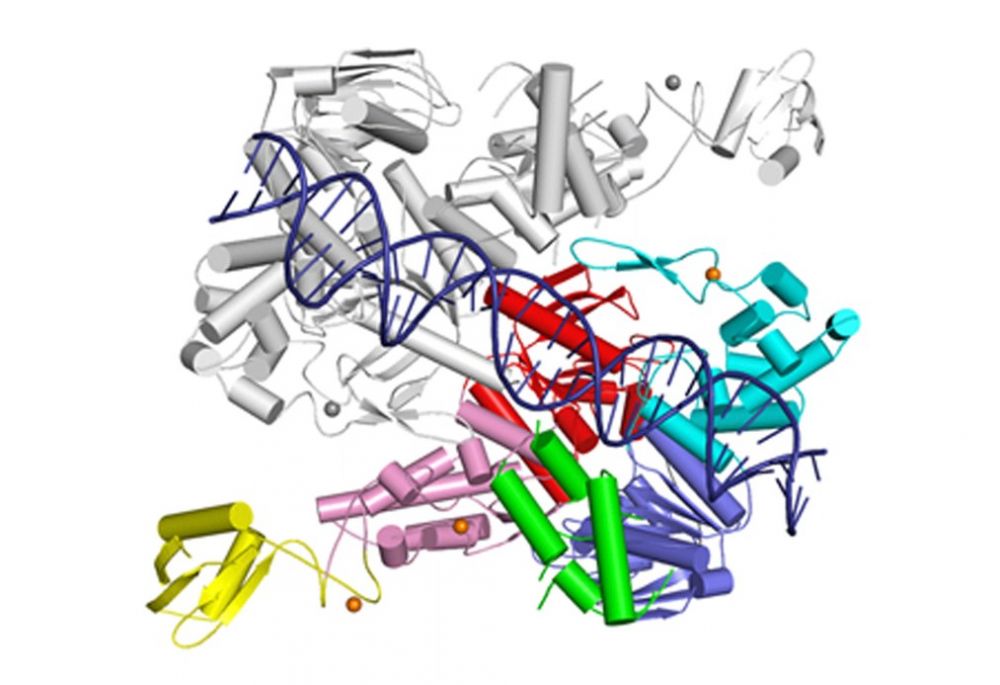
RecFOR
RecFOR pathway is one of the homologous recombination-based DNA repair pathways in bacteria. It involves RecF, RecO and RecR proteins, that bind at the junction of single-stranded (ss) and double-stranded (ds) DNA and then facilitate the replacement of the SSB protein, which initially covers ssDNA, with RecA which promotes the search for the homologous sequence.
Nirwal S, Czarnocki-Cieciura M, Chaudhary A, Zajko W, Skowronek K, Chamera S, Figiel M, Nowotny M. Mechanism of RecF–RecO–RecR cooperation in bacterial homologous recombination. Nature Structural & Molecular Biology, 2023; doi: 10.1038/s41594-023-00967-z.
- The structure of RecF-DNA complex at 3.1 Å resolution, shows that RecF dimer uses its helical protrusions to clamp the dsDNA.
- The structure of RecF-RecR-DNA subcomplex (3.1 Å resolution), shows that one protomer of RecF dimer binds two different regions of the tetrameric RecR ring.
- The structures of RecR-RecO-DNA complex (6.1 Å resolution) and RecF-RecO-RecR-DNA assembly, explains how RecO is positioned to interact with ssDNA and SSB, which is proposed to lock the complex at a ss-dsDNA junction.
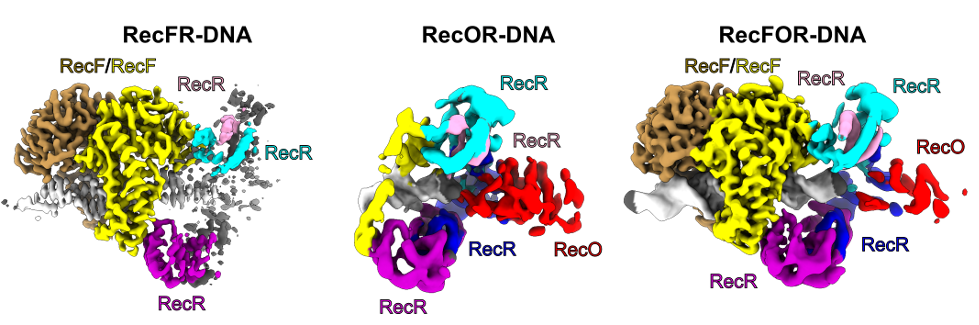
The cryo-EM reconstructions for the RecFOR-DNA assembly and the RecFR-DNA and RecOR-DNA subcomplexes of the assembly. RecF dimer is shown in yellow and sand; RecR tetramer in cyan, pink, purple and blue; RecO in red and DNA in white.
RuvC
RuvC is a canonical bacterial Holliday junction (HJ) resolvase, which functions as a dimer. HJ are four-way DNA structures formed by the exchange of strands between two helices. They are intermediates in homologous recombination, a process which is used to repair dangerous DNA lesions such as double-strand breaks.
Gorecka, KM, Komorowska W and Nowotny M. Crystal structure of RuvC resolvase in complex with Holliday junction substrate. Nucleic Acids Res., 2013; 41(21):9945-55
-
The first crystal structure of RuvC in complex with a DNA substrate and the first substrate complex structure of a cellular resolvase, solved at 3.8 Å resolution.
-
HJ in a novel tetrahedral conformation with two phosphate groups symmetrically located 1 nt from the HJ exchange point interacting with two active sites of RuvC dimer.
-
Novel mode of HJ recognition relative to phage enzymes for which complex crystal structures had been available.

Crystal structure of RuvC in complex with Holliday junction. The two protomers are shown in pink and orange. The DNA is in blue with cleaved phosphates indicated with spheres.
Górecka KM*, Krepl M*&, Szlachcic A, Poznański J, Šponer J, Nowotny M&. RuvC uses dynamic probing of the Holliday junction substrate to achieve sequence specificity and efficient resolution. Nature Commun. 2019; 10(1):4102; *- equally contributing,
& - corresponding authors
-
Mechanism of sequence specificity and cut coordination revealed by a combination of structural biology, biochemistry, and a computational approach.
-
Correct positioning of the substrate for cleavage requires conformational changes within the bound DNA, which are possible only for the cognate sequence.
-
The conformational changes and the relieving of protein-induced structural tension of the DNA facilitates coordination between the two cuts.
-
The unique DNA cleavage mechanism of RuvC demonstrates the importance of high-energy conformational states in nucleic acid readout.
Studies of RuvC mechanism have been done in collaboration with Jiří Šponer (Institute of Biophysics, CAS)
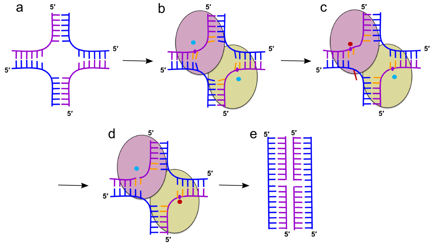
Cartoon representation of the mechanism of HJ resolution by RuvC. (a) Holliday junction. Cleaved and non-cleaved DNA strands are shown in purple and blue ladder-like representations, respectively. (b) Binding of the HJ DNA. The subunits of the dimer are shown as yellow-green and pink ovals. The scissile phosphate is marked as a purple circle. Cyan circles show active sites in an inactive configuration. (c) Flipping of the adenine (red) opposite the scissile base. The active site in the catalytic configuration is shown as a red circle. (d) The second cut. (e) Resolution products
Slx1-Slx4
Slx1 is a nuclease which cleaves various DNA structures during DNA repair and recombination. It associates with Slx4 platform protein which coordinates the action of multiple proteins. Slx1 together with Mus8-Eme1 nuclease constitute one of the major Holliday junction pathways in eukaryotes.
Gaur V, Wyatt HD, Komorowska W, Szczepanowski RH, de Sanctis D, Gorecka KM, West SC, Nowotny M. Structural and Mechanistic Analysis of the Slx1-Slx4 Endonuclease. Cell Rep., 2015; S2211-1247(15)00165-5
-
First structural information for Slx1 and Slx4CCD.
-
Fungal Slx1 forms a homodimer in which the active site is blocked, explaining why Slx1 alone is inactive.
-
Slx4CCD domain binding is mutually exclusive with homodimerization.
-
Slx4 binding exposes the active site of Slx1 and activates the nuclease. This mechanism ensures that the promiscuous and potentially dangerous Slx1 nuclease is only active when bound to Slx4 platform which regulates its activity and coordinates it with other proteins.

Crystal structures of Slx1 homodimer and Slx1 in complex with Slx4CCD domain (orange). Slx1 comprises GIY-IYG nuclease domain (yellow) and RING finger zinc-binding domain (green). Upon Slx4 binding the active site of the nuclease domain is exposed and the enzyme is activated.
Gaur V, Zajko W, Nirwal S, Szlachcic A, Gapińska M, Nowotny M. Recognition and processing of branched DNA substrates by Slx1-Slx4 nuclease. Nucleic Acids Res., 2019; 47:11681-11690
-
Based on a crystal structure, modeling, and biochemical studies, a mechanism was proposed to explain the specificity of Slx1 towards a wide range of branched DNA substrates.
-
Slx1 bends the DNA and identifies the branch point as a flexible discontinuity.
-
Cuts are introduced on the 3ʹ side of the branch point.
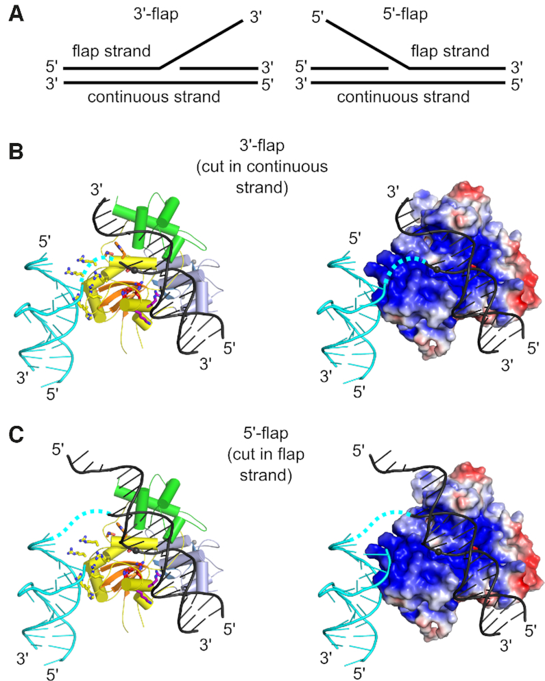
Models of Slx1–Slx4CCD3 interactions with branched DNA. (A) Schematic of the DNA flap substrates with terminology of the strands. (B) Model of Slx1–Slx4CCD3 bound to a 3′-flap substrate in configuration, which is conducive to incision in the continuous strand. (Left) Slx1 GIY-YIG domain is shown in yellow with -strands in orange and RING domain shown in blue. Slx4CCD is shown in green. The modeled DNA is based on the structure of R.Eco29kI restrictase (PDB ID: 3NIC) and is shown in black with the scissile phosphate shown as a sphere. A fragment of the DNA that is observed in the Slx1–Slx4CCD-DNA structure is shown in cyan. The possible link between the two DNA double helices is shown as a dashed cyan line. Residues of the active site are shown as red sticks. The same model with protein in surface representation, colored according to the surface potential (±3 Kt/e). (C) Model of Slx1–Slx4CCD3 bound to a 5′-flap substrate, in configuration which is conducive to incision of the flap strand. The representations are the same as in (B).
RNases H
Bacterial RNase H2
RNase H2 cleaves the 5′ phosphate of ribonucleotides in RNA-DNA junctions. It is the only known enzyme that can initiate the process of mutation-free removal of single ribonucleotides from genomic DNA. Such ribonucleotides are very often misincorporated by replicative polymerases and lead to genomic instability.
Rychlik MP, Chon H, Cerritelli SM, Klimek P, Crouch RJ, Nowotny M. Crystal Structures of RNase H2 in complex with nucleic acid reveal the mechanism of RNA-DNA junction recognition and cleavage. Mol. Cell, 2010; 40:658-670
-
First crystal structure of substrate complex of RNase H2 obtained using T. maritima protein.
-
Unique catalytic mechanism in which the substrate is deformed, so that the phosphate group of the RNA-DNA junction participates in metal ion coordination at the active site. This metal ion positions and activates the nucleophilic water molecule. Hence, the substrate is used to assemble the active site to promote its own cleavage.
The studies of RNases H2 were performed in collaboration with Dr. Robert Crouch (National Institutes of Health, USA).
Crystal structure of T. maritima RNase H2 in complex with the nucleic acid substrate. The catalytic domain is in orange and purple and the helical C-terminal extension in yellow. DNA is shown in blue and single ribonucleotide in red. Metal ions at the active site are shown as green spheres.
Human RNase H2
Human RNase H2 comprises three subunits – the catalytic subunit is very similar to monomeric bacterial and archaeal enzymes while the remaining two subunits do not share sequence similarity with any other known proteins. Mutations in human RNase H2 lead to a severe genetic autoimmune disease - Aicardi-Goutières syndrome.
Figiel M, Chon H, Cerritelli SM, Cybulska M, Crouch RJ, Nowotny M. The structural and biochemical characterization of human RNase H2 complex reveals the molecular basis for substrate recognition and Aicardi-Goutieres syndrome defects. J. Biol. Chem., 2011; 286:10540-50
-
Crystal structure of the trimeric human RNase H2.
-
All mutations from AGS patients mapped and their effect on the structure and mechanism of the enzyme proposed.

Crystal structure of human RNase H2. The catalytic subunit is shown in yellow and the auxiliary ones in purple in blue.
RNase H3
RNases H3 are present in certain bacterial and archaeal species. They share high sequence and structure similarity with RNases H2, however in terms of biochemical properties they are similar to RNases H1 – they prefer to cleave RNA/DNA hybrids in the middle of the RNA sequence. RNase H3 contains a unique N-terminal domain related to TATA-binding protein.
Figiel M, Nowotny M. Crystal structure of RNase H3-substrate complex reveals parallel evolution of RNA/DNA hybrid recognition. Nucleic Acids Res., 2014; 42(14):9285-94
-
The first crystal structure of RNase H3 in complex with the RNA/DNA substrate.
-
The RNA strand is recognized by contacts between 2’-OH groups of four consecutive ribonucleotides.
-
The DNA strand is recognized by deformation to B-form sugar puckers only allowed for DNA.
-
The N-domain specifically binds RNA/DNA hybrid by recognizing 2’-OH of RNA and forming stacking interactions with the ribose rings of the DNA.
-
The mechanism of N-domain is very similar to the structurally unrelated hybrid-binding domain present in the N-terminus of RNases H1. This is a likely case of convergent evolution of RNA/DNA recognition.

Crystal structure of RNase H3 (catalytic domain in yellow and N-domain in green) interacting with an RNA/DNA hybrid (RNA in red and DNA in blue). The cleaved phosphate is shown as a red sphere and the phosphate group of the deformed DNA residue as a blue sphere.
1.
-
May 9, 2021 - Recruitment announcement published
-
May 24, 2021 - Start of the recruitment
-
June 6, 2021 - Deadline for documents submission
Research projects for admissions 2021/2022-4:
-
Poly(A) tails - central hubs of mRNA stability control, Professor Andrzej Dziembowski
-
Signaling of AXL receptor in cancer cells, Professor Marta Miączyńska, Daria Zdżalik-Bielecka, PhD
Poly(A) tails - central hubs of mRNA stability control
Supervisor: Professor Andrzej Dziembowski
Institute: International Institute of Molecular and Cell Biology in Warsaw
Laboratory: Laboratory of RNA Biology
Project description:
Gene expression is regulated at multiple levels. Our lab is interested in the regulation of mRNA stability, especially through the modifications of poly(A) tails. Recently, we have shown that the addition of untemplated uridines to the 3’ end of LINE1 retrotransposons precludes their propagation (Warkocki et al. Cell 2018). Moreover, we have identified a family of poly(A) polymerases TENT5, which reside in the cytoplasm and enhance the expression of mRNAs encoding secreted proteins (Moczek et al. Nature com. 2017; Bilska et al. Nature com. 2020; Gewartowska et al. Cell reports 2021). Those enzymes are differentially expressed in tissues and organs, affecting several aspects of animal physiology. TENT5C is an onco-suppressor in multiple myeloma and control immunoglobulin expression in B cells. TENT5A is essential for collagen secretion, and its mutations lead to congenital bone disease
To study the dynamics of poly(A) tails genome-wide, we have implemented a Direct RNA sequencing Nanopore methodology. It is now widely used in our projects, and we also collaborate with other laboratories interested in post-transcriptional gene expression regulation (for instance, Scheer et al. Nature com. 2021). Moreover, we use Direct RNA sequencing to look globally at the regulation of poly(A) tails (Tudek et al. Nature com., under revision).
In the future, we will continue to study the role and mechanism of action of TENT5 poly(A) polymerases, analyze global control of poly(A) tail lengths and develop bioinformatics tools for Direct RNA sequencing. Finally, we are planning to translate our knowledge out poly(A) tails for the design of mRNAs, which are more stable and better translated, which will be very valuable for mRNA-based therapeutics such as mRNA vaccines.
Aim:
The exact nature of the project will depend on the skills, predispositions, and interests of the selected PhD student. It may focus on:
-
functional analysis of the TENT5A poly(A) polymerases in transgenic mouse models generated in-house using CRISPR/Cas9 methodology
-
mastering of the DRS methodology in either the experimental part or bioinformatic analysis
-
analysis of principles of mRNA stability control and design of more efficient mRNA based therapeutics
Requirements:
-
Master's degree in biology, biochemistry or related field
-
Eligibility for PhD studies in Poland
-
Highly talented individuals who are passionate about research and are full of scientific curiosity
-
Experience in either: molecular biology/transcriptomics, animal models, bioinformatic analysis of transcriptomic data, will be a clear benefit
-
Written and spoken fluency in English
-
Willingness to learn and take new challenges, ability to work independently, analytical thinking
-
Good interpersonal skills and a collaborative attitude.
Number of positions available: 2
Contact: Andrzej Dziembowski
The role of mTOR-Brg1 interaction in normal and aberrant neuronal activity (NCN/MAESTRO)
Supervisor: Professor Jacek Jaworski
Institute: International Institute of Molecular and Cell Biology in Warsaw
Laboratory: Laboratory of Molecular and Cellular Neurobiology
Project description:
Gene expression is key for brain development and function and is regulated by a complex protein apparatus, which, among other things, is responsible for changes in the spatial packing of DNA in the cellular nucleus. mTOR kinase is one of the basic regulators of metabolism Mutations in the mTOR regulating genes, i.e. TSC1 or TSC2 lead to multi-organ diseases with serious neurological and neuropsychological symptoms. One of such diseases is tuberous sclerosis complex characterized by the occurrence of epilepsy, mental retardation and autism spectrum disorders. mTOR acts on many proteins changing their function, but occurs mainly in cytoplasm. However, the results of our previous research and the preliminary data which form the basis of this research proposal indicate that neuronal activity causes mTOR to move to the nucleus of the cell, where it interacts with Brg1. Using advanced molecular, cellular biology and microscopy methods we plan to study how mTOR-Brg1 interaction affects neuronal activity, epileptogenesis and social interactions. The research will use in vitro cultured neurons (rat and human) and Danio rerio. The results will contribute to a better understanding of the role of mTOR in physiology and brain diseases.
Aim:
On the basis of our previous research, we hypothesize that neuronal activity causes mTOR to move to the nucleus, where it regulates the chromatin-modelling complexes and gene expression. At the same time, we hypothesize that this sequence of events is disturbed in tuberous sclerosis complex leading to epilepsy as well as disturbances in social interactions characteristic of autism spectrum diseases. The aim of the project is to verify these hypotheses.
Requirements:
-
Master's degree in biology, biochemistry or related field
-
Eligibility for PhD studies in Poland
-
Good knowledge of basics of molecular and cell biology and/or neurobiology
-
Basic hands-on experience in one of the fields: molecular & cell biology, genetic engineering, fluorescent microscopy, danio rerio animal model
-
Basic programing skills (e.g., R)
-
Written and spoken fluency in English
-
Willingness to learn and take new challenges, ability to work independently, analytical thinking
-
Good interpersonal skills and a collaborative attitude
Number of positions available: 1
Contact: Jacek Jaworski
Signaling of AXL receptor in cancer cells
Supervisor: Professor Marta Miączyńska, auxiliary supervisor Daria Zdżalik-Bielecka, PhD
Institute: International Institute of Molecular and Cell Biology in Warsaw
Laboratory: Laboratory of Cell Biology
Project description:
AXL is a tyrosine kinase receptor that upon activation by its GAS6 ligand regulates cell growth, survival, migration and invasion. Overexpression of AXL occurs in a wide variety of cancer types
and is associated with increased invasiveness and metastasis, as well as resistance of cancer cells to anticancer therapies. In our previous studies, we identified the AXL receptor interactome (a set of proteins with which the receptor interacts) and discovered that AXL binds numerous proteins that regulate actin cytoskeleton dynamics. Through such interactions, activation of AXL receptor stimulates the formation of peripheral and circular membrane ruffles that promote macropinocytosis and ultimately lead to cell invasion (see our manuscript at https://bit.ly/2QfEVpF for more details). Macropinocytosis is a form of endocytosis that allows cells to take up large volumes of extracellular fluid and the compounds dissolved within it. The macromolecules taken up through this pathway can be an important source of nutrients that fuel cancer cell growth. It has been recently proposed that macropinocytosis may also contribute to resistance of cancer cells to chemotherapeutics. We found that proteins in the AXL interactome may be involved in nutrient uptake by macropinocytosis. Elucidating their mechanisms of action in AXL receptor signaling will be the goal of the proposed project.
Aim:
The goal of this project is to investigate the molecular mechanisms of AXL receptor signaling that regulates cancer cell growth. Specifically, we will investigate how the AXL receptor and its ligand GAS6 stimulate uptake of nutrients from the extracellular environment via macropinocytosis.
We will also determine whether and how this phenomenon contributes to drug resistance of cancer cells and, consequently, to the growth of tumors despite the applied therapy.
Requirements:
-
Master's degree in biology, biochemistry or related field
-
Eligibility for PhD studies in Poland
-
Solid understanding of the principles of molecular and cell biology
-
Previous experience in laboratory work and familiarity with basic molecular biology techniques
-
Written and spoken fluency in English
-
Good interpersonal skills and a collaborative attitude
Number of positions available: 1
Contact: This email address is being protected from spambots. You need JavaScript enabled to view it., Daria Zdżalik-Bielecka
RNA-Protein Interactions in Human Health and Disease (NCN/DIOSUCRI)
Supervisor: Gracjan Michlewski, PhD, Professor
Institute: International Institute of Molecular and Cell Biology in Warsaw
Laboratory: Laboratory of RNA-Protein Interactions
Project description:
RNA-binding proteins (RBPs) are key molecules that control gene expression through RNA-protein interactions. Consequently, they contribute to cellular homeostasis, normal development and majority of human diseases. Importantly, new RBPs are being discovered by high-throughput proteomics, but we still have a limited understanding of their function.
RNA viruses have caused several epidemics in the 21st century. Taking influenza A virus (IAV) infection as an exemplar, it kills 250,000 to 500,000 people annually and generates a significant global socioeconomic burden. Importantly the emergence of COVID-19 pandemic caused by an RNA virus SARS-CoV-2 continue to have catastrophic consequences on public health and world economy. Thus, a detailed molecular understanding of host-virus interactions is imperative in order to know how best to inactivate these viruses and prevent major disruptions in the future.
We have recently discovered and started characterising novel RNA binding protein – E3 ubiquitin ligase TRIM25 (Choudhury et al. 2014; Choudhury et al. 2017). TRIM25 belongs to a large family of tripartite motif-containing proteins (more than 80), most of which have E3 ubiquitin ligase activity. Many of TIRIMs are positive or negative regulators of innate immune response pathways. Importantly, TRIM25 is emerging as a key factor in the innate immune response to RNA viruses (including IAV, CoV, dengue virus and many others). Despite the essential involvement of TRIM25 in viral RNA-induced innate immunity, its RNA-binding functions are still poorly understood.
Aim:
With this project, we aim to take advantage of an assembled multi-disciplinary team to uncover the roles of the novel RNA-protein interactions in the antiviral response to selected RNA virus infections. We hypothesise that TRIM25 binds directly to viral RNAs to restrict virus propagation. We also hypothesise that other members from TRIM family bind RNA. Finally, we hypothesise that specific host RBPs bind to virus derived RNAs and inhibit or augment innate immune response. In summary, this project has the potential to make crucial contributions to understanding the innate immune response to RNA viruses and provide a platform for the development of novel, RNA-based antiviral therapeutics.
Requirements:
-
Master's degree in biology, biochemistry or related field
-
Solid knowledge of the principles of cell and molecular biology, virology or biochemistry
-
Hands-on experience in laboratory work and is familiar with basic cell and molecular biology techniques
-
Prior experience in virus handling and analysis, cell culture, mass spectrometry or bioinformatics will be an advantage
-
Proficiency in written and spoken English
-
Excellent interpersonal skills, initiative and ability to work independently and in a high-performance team
Number of positions available: 1
Contact: Gracjan Michlewski
Identifying unique adaptive responses of red pulp macrophages to iron deficiency (NCN/SONATA)
Supervisor: Wojciech Pokrzywa, PhD DSc, auxiliary supervisor, PI: Katarzyna Mleczko-Sanecka, PhD
Institute: International Institute of Molecular and Cell Biology in Warsaw
Laboratory: Laboratory of Iron Homeostasis
Iron deficiency is a global health burden with profound socio-medical impacts, but little is known about how functions of specialized cells are affected by low systemic iron levels. Red pulp macrophages (RPMs) residing in the spleen are responsible for removing aged erythrocytes from the bloodstream. Following erythrocyte lysis, RPMs release iron to the circulation to replenish the pool of serum iron necessary for sustaining erythropoiesis. RPMs are thus critical for maintaining blood and iron homeostasis in the body. Interestingly, it was largely unknown if and how key cellular functions of RPMs are affected by low body iron status. Using a mouse model of nutritional iron deficiency, we uncovered that iron deficiency triggers specific but still elusive signaling mechanisms that modulate RPMs’ phagocytic and metabolic functions. We expect that these newly identified responses likely contribute to the adaptation of the whole organism to limited iron supplies. Within our project, we apply both in vivo and in vitro approaches to decipher the molecular mechanism responsible for the adaptive functional ‘rewiring’ of RPMs in iron deficiency and determine their physiological role for the whole organism. The new knowledge generated by this research is expected to significantly advance our understanding of an organism’s adaptation to iron deficiency.
Aim:
We aim to comprehensively characterize the functional adaptation of RPMs to iron deficiency and identify its molecular triggers. We also plan to determine how this ‘rewiring’ affects RPMs’ inflammatory status. We will explore how the abrogation of this ‘adaptation program’ affects the organism, including iron and blood homeostasis. To this end, we will create and characterize new conditional knock-out mice characterized by specific suppression of the RPMs’ adaptation to low iron conditions.
Requirements:
-
Master's degree in biology, biochemistry or related field
-
Eligibility for PhD studies in Poland
-
Interests in molecular aspects of physiology, motivation for experimental work, passion for science, hands-on experience in laboratory work
-
Experience in mouse/rat-based studies or willingness to work with animals
-
Written and spoken fluency in English
-
Willingness to learn and take new challenges, ability to work independently, analytical thinking
-
Good interpersonal skills and a collaborative attitude.
-
Research achievements (eg, publications or manuscripts in preparation) and experience abroad will be of advantage
Number of positions available: 2
Contact: This email address is being protected from spambots. You need JavaScript enabled to view it.
2.
-
August 4, 2021 - Start of the recruitment
-
August 18, 2021 - Deadline for documents submission
Research projects for admissions 2021/2022-5:
-
Genomics and Epigenomics of acute myelogenous leukemia (AML), Professor Matthias Bochtler
-
Poly(A) tails - central hubs of mRNA stability control, Professor Andrzej Dziembowski
Genomics and Epigenomics of acute myelogenous leukemia (AML)
Supervisor: Professor Matthias Bochtler
Institute: International Institute of Molecular and Cell Biology in Warsaw
Laboratory: Laboratory of Structural Biology
Epigenomic changes play a prominent role in acute myelogenous leukemia (AML). Mutations in the methyltransferase DNMT3A and the dioxygenase TET2 are among the most frequent alterations in this type of malignancy. It has been proposed that defects in epigenomics entail DNA repair defects, which in turn lead to karyotype degradation. This contrasts with information from the Cancer Genome Atlas and the COSMIC database, which both identify AML as a typical M-type malignancy, i.e. a malignancy that is driven by mutations, rather than by copy-number variation. However, clinical observation suggests that a considerable fraction of AML patients have karyotype aberrations. In some cases, the chromosomal changes can be drastic and resemble the chromothripsis seen in other malignancies. Highly karyotype aberrant AMLs are poorly characterized. It is not clear whether the spectrum of exome mutations is similar in these leukemias and in M-type leukemias and what drives the karyotype degradation. It is also unclear whether changes in the epigenomic machinery and their possible effects on DNA signaling play a role in this process. We hope to clarify these issues, primarily by sequencing approaches, in collaboration with clinicians in Heidelberg and Dresden (Germany).
Aim
The aim of the project is to obtain deep sequencing data for AML patients (bulk and single cell). We plan to compare the spectrum of mutations and copy number variation in highly aberrant and normal karyotype AMLs. We plan to test if the spectrum of mutations in the coding genome and tumor clonal histories are similar in the two types of AML. We want to learn if mutations in the epigenomic enzymes cause DNA repair phenotypes, which could ultimately lead to karyotype degradation.
Requirements:
-
Master's degree in biology, biochemistry or a related field
-
Eligibility for PhD studies in Poland
-
Theoretical knowledge of genetics and epigenetics.
-
Practical experience with cellular fractionation by FACS.
-
Experience with preparation of libraries for DNA sequencing using Nanopore and Illumina technologies.
-
Experience with genotyping (high resolution melting analysis).
-
Experience with or at least interest in bioinformatic analysis of deep sequencing data.
-
Written and spoken fluency in English
-
Willingness to learn and take new challenges, ability to work independently, analytical thinking
-
Good interpersonal skills and a collaborative attitude
Number of positions available: 1
Contact: This email address is being protected from spambots. You need JavaScript enabled to view it., This email address is being protected from spambots. You need JavaScript enabled to view it.
Poly(A) tails - central hubs of mRNA stability control
Supervisor: Professor Andrzej Dziembowski
Institute: International Institute of Molecular and Cell Biology in Warsaw
Laboratory: Laboratory of RNA Biology
Project description:
Gene expression is regulated at multiple levels. Our lab is interested in the regulation of mRNA stability, especially through the modifications of poly(A) tails.
Recently, we have shown that the addition of untemplated uridines to the 3’ end of LINE1 retrotransposons precludes their propagation (Warkocki et al. Cell 2018). Moreover, we have identified a family of poly(A) polymerases TENT5, which reside in the cytoplasm and enhance the expression of mRNAs encoding secreted proteins (Moczek et al. Nature com. 2017; Bilska et al. Nature com. 2020; Gewartowska et al. Cell reports 2021). Those enzymes are differentially expressed in tissues and organs, affecting several aspects of animal physiology. TENT5C is an onco-suppressor in multiple myeloma and control immunoglobulin expression in B cells. TENT5A is essential for collagen secretion, and its mutations lead to congenital bone disease.
To study the dynamics of poly(A) tails genome-wide, we have implemented a Direct RNA sequencing Nanopore methodology. It is now widely used in our projects, and we also collaborate with other laboratories interested in post-transcriptional gene expression regulation (for instance, Scheer et al. Nature com. 2021). Moreover, we use Direct RNA sequencing to look globally at the regulation of poly(A) tails (Tudek et al. Nature com., under revision).
In the future, we will continue to study the role and mechanism of action of TENT5 poly(A) polymerases, analyze global control of poly(A) tail lengths and develop bioinformatics tools for Direct
RNA sequencing. Finally, we are planning to translate our knowledge out poly(A) tails for the design of mRNAs, which are more stable and better translated, which will be very valuable for mRNA-based therapeutics such as mRNA vaccines.
Aim:
The exact nature of the project will depend on the skills, predispositions, and interests of the selected PhD student. It may focus on:
-
functional analysis of the TENT5A poly(A) polymerases in transgenic mouse models generated in-house using CRISPR/Cas9 methodology.
-
mastering of the DRS methodology in either the experimental part or bioinformatic analysis.
-
analysis of principles of mRNA stability control and design of more efficient mRNA based therapeutics
Requirements:
-
Master's degree in biology, biochemistry or related field
-
Eligibility for PhD studies in Poland
-
Highly talented individuals who are passionate about research and are full of scientific curiosity
-
Experience in either: molecular biology/transcriptomics, animal models, bioinformatic analysis of transcriptomic data, will be a clear benefit
-
Written and spoken fluency in English
-
Willingness to learn and take new challenges, ability to work independently, analytical thinking
-
Good interpersonal skills and a collaborative attitude
Number of positions available: 2
Contact: This email address is being protected from spambots. You need JavaScript enabled to view it.
Experimental analysis of molecular determinants involved in epilepsy (NCN/OPUS)
Supervisor: Professor Jacek Kuźnicki, auxiliary supervisor/PI: Vladimir Korzh, PhD
Institute: International Institute of Molecular and Cell Biology in Warsaw
Laboratory: Laboratory of Neurodegeneration
Project description:
The effects of KCNB1 mutations that cause epileptic encephalopathy were analyzed mechanistically mainly using electrophysiology in heterologous cells in vitro. The developmental analysis was limited by the availability of single mutants in mice and zebrafish and did not explore the whole range of effects caused by KCNB1 mutations (Shen et al., 2016). The KCNB1 loss of function (LOF) or gain of function (GOF) cause specific morphological changes in the brain ventricles (Shen et al., 2016) and inner ear (Jedrychowska et al., 2020) in developing zebrafish embryos and larvae. The zebrafish transgenics express fluorescent markers in specific manner. The high-resolution microscopy of transgenic embryos and larvae in vivo provides information about developmental mechanisms as well as changes in activity of specific signaling pathways. These tools satisfy conditions necessary to study the developmental effect of different KCNB1 mutations in real time. This rationale was confirmed in preliminary experiments when analyzing the effect of overexpression of human mutated KCNB1 mRNA. Kcnb1 GOF causes cell delamination in brain ventricles and their expansion (hydrocephalus), and enlargement of the inner ear and otoliths. These features recapitulate the phenotype of the kcnb1 GOF zebrafish mutant and constitute the rationale for the "brain and ear" in vivo test to be used when defining an effect of human mutations.
Aim:
Using the site-specific CRISPR-Cas9 mutagenesis in zebrafish, the representative Kcnb1 mutations that mimic the known human mutations of KCNB1 will be generated and analyzed by a combination of bioimaging, single-cell transcriptomics, electrophysiology and behavioral analysis. This will provide rationale for subfunctionalization of human KCNB1 mutations.
Requirements:
-
Master's degree in biology, biochemistry or related field
-
Eligibility for PhD studies in Poland
-
Prior experience in molecular developmental biology and zebrafish studies is a bonus during selection of candidates, but necessary training will be provided
-
Written and spoken fluency in English
-
Willingness to learn and take new challenges, ability to work independently, analytical thinking
-
Good interpersonal skills and a collaborative attitude
Number of positions available: 2
Contact: Vladimir Korzh
Identifying unique adaptive responses of red pulp macrophages to iron deficiency (NCN/SONATA)
Supervisor: Wojciech Pokrzywa, PhD DSc, auxiliary supervisor, PI: Katarzyna Mleczko-Sanecka, PhD
Institute: International Institute of Molecular and Cell Biology in Warsaw
Laboratory: Laboratory of Iron Homeostasis
Project description:
Iron deficiency is a global health burden with profound socio-medical impacts, but little is known about how functions of specialized cells are affected by low systemic iron levels. Red pulp macrophages (RPMs) residing in the spleen are responsible for removing aged erythrocytes from the bloodstream. Following erythrocyte lysis, RPMs release iron to the circulation to replenish the pool of serum iron necessary for sustaining erythropoiesis. RPMs are thus critical for maintaining blood and iron homeostasis in the body. Interestingly, it was largely unknown if and how key cellular functions of RPMs are affected by low body iron status. Using a mouse model of nutritional iron deficiency, we uncovered that iron deficiency triggers specific but still elusive signaling mechanisms that modulate RPMs’ phagocytic and metabolic functions. We expect that these newly identified responses likely contribute to the adaptation of the whole organism to limited iron supplies. Within our project, we apply both in vivo and in vitro approaches to decipher the molecular mechanism responsible for the adaptive functional ‘rewiring’ of RPMs in iron deficiency and determine their physiological role for the whole organism. The new knowledge generated by this research is expected to significantly advance our understanding of an organism’s adaptation to iron deficiency.
Aim:
We aim to comprehensively characterize the functional adaptation of RPMs to iron deficiency and identify its molecular triggers. We will explore how the abrogation of this ‘adaptation program’ affects the organism, including iron and blood homeostasis. To this end, we will create and characterize new conditional knock-out mice characterized by specific suppression of the RPMs’ adaptation to low iron conditions.
Requirements:
-
Master's degree in biology, biochemistry or related field
-
Eligibility for PhD studies in Poland
-
Interests in molecular aspects of physiology, motivation for experimental work, passion for science, hands-on experience in laboratory work
-
Experience in mouse/rat-based studies or willingness to work with animals
-
Written and spoken fluency in English
-
Willingness to learn and take new challenges, ability to work independently, analytical thinking
-
Good interpersonal skills and a collaborative attitude.
-
Research achievements (eg, publications or manuscripts in preparation) and experience abroad will be of advantage
Number of positions available: 1
Contact: Katarzyna Mleczko-Sanecka
3.
Research projects for admissions 2021/2022-6:
9.1 Poly(A) tails - central hubs of mRNA stability control (prof. Andrzej Dziembowski)9.1 Poly(A) tails - central hubs of mRNA stability control (prof. Andrzej Dziembowski) - 2 places available
9.3 Ten eleven translocation 2 (TET2) in acute myeloid leukemia (prof. Matthias Bochtler)
1.
-
May 25, 2020 - recruitment opens
-
June 7, 2020 - deadline for applications
Research projects for admissions 2020/2021-1:
Linking abnormal Ca2+ signaling and the unfolded protein response with Huntington’s disease pathology in both YAC128 mouse model and iPSC-derived neurons from HD patients.
Prof. Jacek Kuźnicki, PhD; Magdalena Czeredys, PhD, Laboratory of Neurodegeneration
Description: Huntington’s disease (HD) is a progressive neurodegenerative disorder characterized by the aggregation of mutant huntingtin and degeneration of medium spiny neurons (MSNs) in the striatum. Abnormal Ca2+ signaling is considered as an early event in HD pathology since disturbances in Ca2+ homeostasis were found in HD models and postmortem samples from HD patients. One of the pathways for Ca2+ signaling is store-operated calcium entry (SOCE). The activation of inositol-(1,4,5)triphosphate receptor 1 (IP3R1) results in Ca2+ release, which decreases ER Ca2+ content and activates Ca2+ influx through SOC channels. Elevated SOCE and increased IP3R1 activity was previously reported in MSNs from the transgenic model of HD, YAC128. The project is based on the hypothesis that neurodegeneration in HD is induced by disturbances in Ca2+ signaling in neurons. Previously we found that huntingtin-associated protein 1 (HAP1) that is overexpressed in striatal neurons and binds to mutant huntingtin causes dysregulation of Ca2+ signaling by increased activation of both SOCE and IP3R1 receptors. We intend to examine the link between dysregulated Ca2+ signals and neuronal cell death in HD. The experiments will be performed in YAC128 MSNs cultures and neurons delivered by the reprogramming of fibroblasts from HD patients with the application on CRISPR/Cas9-based editing strategies and Ca2+ signaling inhibitors.
Aim: The project aims to investigate whether and how the disturbed Ca2+ homeostasis affects HD pathology. A Ph.D. project related to this issue will be done using different HD models. One position is available in the project. We are looking for a person interested in neurobiology, with experience in working with animal models (mice, zebrafish), cell cultures, and biochemical techniques (immunoprecipitation, western blot). Knowledge/experience in iPSCs cultures is welcome.
Number of positions available: 1
Source of funding: NCN/OPUS grant
Contact: This email address is being protected from spambots. You need JavaScript enabled to view it.
Cytoplasmic polyadenylation as a central regulator of physiological processes
Prof. Andrzej Dziembowski, PhD, Laboratory of RNA Biology
Description: Poly(A) tails generated by canonical poly(A) polymerases during mRNA 3’ formation are essential for mRNA stability and translation. It is now appreciated that poly(A) tail dynamics is more complex than previously suspected; deadenylated mRNAs in the cytoplasm can be degraded, uridylated or stored in a dormant state to be later re adenylated to activate protein synthesis. Cytoplasmic polyadenylation was mostly studied in the context of gametogenesis and in synapses, where the transcriptional activity is limited. Surprisingly, mouse lines devoid of the well known cytoplasmic poly(A) polymerase GLD2 display no apparent phenotypes. We recently described a novel family of cytoplasmic poly(A) polymerases, TENT5 (FAM46), comprising four members in vertebrates (Mroczek et al. & Bilska et al. Nat Comm 2017,2020). TENT5C acts as a tumor suppressor in multiple myeloma, while mutations in TENT5A lead to a rare disease osteogenesis imperfecta. We have generated KO mouse models for all TENT5 genes and detected a variety of different phenotypes affecting several organs and biological processes: gametogenesis, growth, skeletal development, hematopoiesis, immune response, and behavior. Moreover, analysis of the KO of worm TENT5 orthologue revealed dysfunction of innate immunity. Thus, TENT5 proteins contribute significantly to metazoan physiology and, more generally, that cytoplasmic polyadenylation plays a much broader role than previously anticipated, opening a new area of important research.
Aim: The project aims is to dissect functions and mechanisms of cytoplasmic polyadenylation by TENT5 in innate immunity, erythropoiesis, and neuronal physiology. Unique animal models constructed using CRISPR/Cas9, combined with advanced transcriptomic and proteomic approaches, will be used to achieve our goals. Several positions are available in the lab. The exact PhD project will depend on the particular skills and preferences of the student. We are looking for students with experience in work with animal modes (mouse, C. elegans), RNA biology, or bioinformatics.
Number of positions available: 4
Source of funding: NCN/Norway grants/FNP/Horizon2020 era chairs
Contact: This email address is being protected from spambots. You need JavaScript enabled to view it.
See also:
2.
-
05/08 - recruitment opens
-
18/08 - deadline for applications
-
7/09 – 17/09 - interviews
-
21/09 – publication of results
Research projects for admissions 2020/2021-2:
Title: Elucidating the epigenetic contribution to cardiovascular lineage specification
Supervisor: Cecilia Winata
Institute: International Institute of Molecular and Cell Biology in Warsaw
Laboratory: Laboratory of Zebrafish Developmental Genomics
Background:
One key question in the field of organogenesis relates to how the many types of cells required to make an organ are generated from a pool of progenitor cells with initially similar characteristics. Heart (cardiac) progenitor cells are located close to those that will also generate blood and blood vessels (hemoangiogenic). At very early stages of embryonic development, they express nkx2.5 in common. Subsequently, each type of cells expresses different sets of genes and adopts epigenetic states which signifies their identity. Despite the knowledge that nkx2.5-expressing progenitors could contribute to diverse cell lineage types, several key questions remain unanswered. First, it is unclear at which developmental stage the segregation between cardiac and hemoangiogenic fates begin to occur. Second, the exact pathway and intermediate stages which these progenitors go through during the process of lineage specification are still largely unknown. In addition, despite the knowledge that cardiac transcription factors including Nkx2.5 itself are known to interact with chromatin modifying factors and promote chromatin changes, it is still unknown to what extent epigenetics play a role in driving cell fate decisions at the individual cell level. By tracing the evolution of cellular heterogeneity over time, and at the same time assessing the dynamics of epigenetic landscape at the single cell level, we will elucidate the mechanism of cardiovascular lineage specification.
Aim:
The goal of this project is to elucidate the epigenetic contribution towards the lineage decision of nkx2.5-expressing progenitors into either cardiac or hemoangiogenic lineage. We hypothesize that distinct epigenetic states occur among subpopulations of nkx2.5-expressing progenitors according to their lineage diversification potential. We will profile open chromatin regions at single cell level anddetermine whether Nkx2.5 plays a role in establishing the epigenetic state by scATAC-seq method(Jia et al., 2018,Nat Commun9, 4877).
Requirements:
-
Bachelor’s or Master’s degree in Biology, Biochemistry, or equivalent
-
Solid understanding of the principles of molecular biology and genetics
-
Previous laboratory experience in molecular biology and/or biochemistry techniques
-
Prior experience in flow cytometry, NGS, and/or working with animal models (mouse or zebrafish), as well as basic programming skills would be an advantage although not essential
-
Ability to communicate fluently in English and has a collaborative attitude
Title: Lysine deserts as a universal mechanism to escape premature proteins degradation
Supervisor: dr hab. Wojciech Pokrzywa
Institute: International Institute of Molecular and Cell Biology in Warsaw
Laboratory: Laboratory of Protein Metabolism in Development and Aging
Background:
Eukaryotic cells use degradation systems such as the ubiquitin-proteasome (UPS) system to remove unwanted proteins. UPS mediates proteolysis by attaching the small protein ubiquitin to the target protein, using a cascade of enzymes in a process called ubiquitination. Ubiquitin is attached primarily to a specific amino acid in the target protein - lysine. Recent studies have shown that the yeast protein Slx5 avoids UPS due to the extensive, lysine-free region in the so-called lysine desert. Such a lysine desert may be a part of the uncharacterized strategy used by proteins to avoid premature degradation. As a result of our initial analyzes, we found various lysine deserts among the proteins of both simple and complex organisms. Many of these proteins are associated with the UPS, suggesting a protective mechanism against self-determination for degradation. However, we have also found lysine deserts in proteins involved in fundamental cellular processes such as transcription.
Aim:
The overall objective of these studies is to understand the common occurrence of proteins with lysine deserts using evolutionary and structural bioinformatics analyzes supported by experimental methods. The theoretical part will consist of a quantitative analysis of the evolutionary preservation of lysine deserts in a number of taxonomic groups, followed by a qualitative analysis of their biological functions. Selected proteins will also be analyzed via molecular simulations to understand the dynamics of lysine desert exposure under selected conditions. The most promising candidates obtained from bioinformatics analyzes will be subjected to experimental tests using human cell lines and animal model Caenorhabditis elegans.
Requirements:
-
Master's degree in biological sciences/bioinformatics
-
experience in programming (Python/R), omics data analysis and cluster computing
-
knowledge of biological databases, biochemistry, molecular biology, evolutionary and structural bioinformatics of proteins
-
fluency in English
-
strong motivation for scientific work (documented internships and apprenticeships in scientific institutes)
-
ability to organize working time independently
-
systematic work
-
experience in molecular modeling and/or laboratory work/C. elegans maintenance is an advantage
Title: The impact of cytoplasmic polyadenylation on local translation in neurons
Supervisor: prof. dr hab. Andrzej Dziembowski
Institute: International Institute of Molecular and Cell Biology in Warsaw
Laboratory: Laboratory of RNA Biology
Background:
Neurons communicate with each other through synapses, specialized contact sites that enable electrical impulses to be transmitted between cells. Synapses are small, but partly independent compartments of the neuron because they contain the molecular machinery indispensable for protein synthesis. This process of protein production on the basis of mRNAs transported to distant synapses from the cell body is called local translation. Local protein synthesis is essential for the proper functioning of the synapse, and its dysregulation is the cause of severe neurodevelopmental disorders. In recent years, thanks to the development of new technologies, we have learned more about these essential processes taking place in synapses. However, the precise molecular mechanisms by which synaptic translation is regulated is still far from being understood.
The ends of mRNA molecules are specifically modified in order to enhance their stability and ability to serve as a template for proteins synthesis at ribosomes: at the 5’ end so-called cap structure is positione, while at the end, there is a poly(A) tail. Nearly all mRNAs in the cell are polyadenylated in the nucleus right after being transcribed from DNA and before their transport to the cytoplasm. However, there is growing evidence that the process of polyadenylation can also take place in the cytoplasm and is therefore called cytoplasmic polyadenylation. In neurons, cytoplasmic polyadenylation of synaptic mRNAs plays a significant role in the regulation of protein synthesis. However, until now, it was studied only for a few mRNAs, and the global impact of this phenomenon and the specific enzymes carrying out the reactions are unknown.
This research project will be performed in cooperation with Prof. Clive Bramham from the University of Bergen and Prof. Magdalena Dziembowski from CENT UW.
Aim:
We aim to elucidate the function and mechanism of cytoplasmic polyadenylation of neurons. To achieve our goals, unique animal models constructed using the CRISR/Cas9 method, combined with advanced transcriptomic and proteomic approaches, will be used.
Two positions are available in the laboratory. The exact project will depend on the specific skills and preferences of the student. We are looking for students with experience in working with animal models, RNA biology or bioinformatics.
See also:
3.
-
December 8, 2020 - Recruitment announcement published
-
December 22, 2020 - Start of the recruitment
-
January 5, 2021 - Deadline for documents submission
Research projects for admissions 2020/2021-3:
1. Title: Rac1 contribution to brain connectivity impairments and neuropsychiatric disorders in Tuberous Sclerosis Complex (NCN/OPUS)
Supervisor: Prof. Jacek Jaworski
Auxiliary Supervisor: Justyna Zmorzyńska, PhD
Institute: International Institute of Molecular and Cell Biology in Warsaw
Laboratory: Laboratory of Molecular and Cellular Neurobiologylular Neurobiology
During the brain development specific cellular events, including establishment of neural polarity, axon elongation, and synapse formation, happen in a temporally and spatially controlled manner to establish connectivity. These temporal and spatial boundaries are created by tight regulation of intrinsic and extrinsic factors. One of major components that may perform this regulation during connectivity development is Rac1 as it links plasma membrane receptors with actin dynamics. Also, mammalian target of rapamycin (mTOR) integrates intra- and extracellular factors. The diseases associated with over-expression of mTOR usually exhibit impairments of the brain development and neuropsychiatric phenotypes that do not necessarily correlate with mutation burden or cannot necessarily be explained by levels of mTOR activation. One of the examples is Tuberous Sclerosis Complex (TSC) in which neuropsychiatric disorders like autism spectrum disorder, intellectual disability, or anxiety do not fully correlate with mTOR expression levels, epilepsy, or tumor burden. Therefore, other molecular pathways must interact with mTOR pathway in order to produce these phenotypes. Our results and preliminary data suggest that Rac1 pathway and its diverse inputs may participate in the regulation of neuropsychiatric disorders in TSC through control of connectivity formation during development. The project will be conducted using zebrafish TSC model.
Aim:
The main objective of this project is to unravel how the interplay between various pathways that converge on Rac1 may participate in the brain connectivity and underlie TSC-associated neuropsychiatric disorders. The project will include behavioral testing, extensive microscopy imaging and image analysis, and transcriptome analysis.
Requirements:
-
Master’s degree in Biology, Biochemistry, Bioinformatics, or related area
-
Solid understanding of the principles of molecular and cellular biology; knowledge on brain development, neuroscience, or computer vision will be a plus
-
Willingness to work with zebrafish animal model
-
Previous laboratory experience in basic molecular biology, biochemistry techniques, and/or imaging experience
-
Prior experience in NGS, and/or working with animal models (mouse or zebrafish), as well as basic programming skills would be an advantage although not essential
-
Ability to communicate fluently in English and a collaborative attitude
Number of positions available: 1
Contact: This email address is being protected from spambots. You need JavaScript enabled to view it.
2. Title: Identification of novel vulnerabilities of VPS4B-deficient cancer cells (NCN/OPUS)
Supervisor: Prof. Marta Miączyńska
Auxilliary Supervisor: Ewelina Szymańska, PhD
Institute: International Institute of Molecular and Cell Biology in Warsaw
Laboratory: Laboratory of Cell Biology
Project description:
One of the key challenges in oncology is to effectively kill cancer cells while leaving healthy cells intact. To meet this goal, precision oncology aims to tailor anti-tumor therapies to individual genetic changes in cancer cells of a given patient. To provide new targets for precision oncology we need to understand the relationship between the genetic alterations of cancer cells and the dependencies (vulnerabilities) they cause.
VPS4A and VPS4B enzymes together with the Endosomal Sorting Complex Required for Transport (ESCRT) machinery are involved in membrane remodeling during e.g. endocytosis, cell division, and plasma membrane repair. In our previous project, we identified VPS4B deficiency as a selective weakness of colorectal cancer cells with chromosome 18q deletion. We also demonstrated that survival of VPS4B-depleted cancer cells depends on the presence of VPS4A and characterized the molecular consequences of simultaneous depletion of VPS4 proteins that lead to cell death (more details in Szymańska et al, EMBO Mol Med, 2020). Very probably, VPS4B deficiency makes cancer cells more vulnerable not only to VPS4A loss but also to other perturbations affecting gene(s) cooperating with VPS4B in cellular processes essential for life.
Aim:
We aim to identify and characterize novel vulnerabilities of VPS4B-deficient cancer cells among candidates selected from datasets of the Cancer Dependency Map Project (Broad Institute). To this end, we will examine the impact of simultaneous depletion of VPS4B and a selected candidate on in vitro and in vivo growth of cancer cells. Further, we will elucidate the consequences of depletion of VPS4B and a candidate for cancer-relevant cellular processes, e.g. endocytosis, cytokinesis and migration.
Requirements:
-
Master's degree in biology, biochemistry or related field.
-
Solid understanding of the principles of cell and molecular biology.
-
Previous experience in laboratory work and familiarity with basic molecular biology techniques.
-
Written and spoken fluency in English.
-
Good interpersonal skills and a collaborative attitude.
Number of positions available: 1
Contact: This email address is being protected from spambots. You need JavaScript enabled to view it.
3. RNA-Protein Interactions in Human Health and Disease (NCN/DIOSUCRI)
Supervisor: Gracjan Michlewski, PhD, DSc
Institute: International Institute of Molecular and Cell Biology in Warsaw
Laboratory: Laboratory of RNA-Protein Interactions
Project description:
RNA-binding proteins (RBPs) are key molecules that control gene expression through RNA-protein interactions. Consequently, they contribute to cellular homeostasis, normal development and majority of human diseases. Importantly, new RBPs are being discovered by high-throughput proteomics, but we still have a limited understanding of their function.
RNA viruses have caused several epidemics in the 21st century. Taking influenza A virus (IAV) infection as an exemplar, it kills 250,000 to 500,000 people annually and generates a significant global socioeconomic burden. Importantly the emergence of COVID-19 pandemic caused by an RNA virus SARS-CoV-2 continue to have catastrophic consequences on public health and world economy. Thus, a detailed molecular understanding of host-virus interactions is imperative in order to know how best to inactivate these viruses and prevent major disruptions in the future.
We have recently discovered and started characterising novel RNA binding protein – E3 ubiquitin ligase TRIM25 (Choudhury et al. 2014; Choudhury et al. 2017). TRIM25 belongs to a large family of tripartite motif-containing proteins (more than 80), most of which have E3 ubiquitin ligase activity. Many of TIRIMs are positive or negative regulators of innate immune response pathways. Importantly, TRIM25 is emerging as a key factor in the innate immune response to RNA viruses (including IAV, CoV, dengue virus and many others). Despite the essential involvement of TRIM25 in viral RNA-induced innate immunity, its RNA-binding functions are still poorly understood.
Aim:
With this project, we aim to take advantage of an assembled multi-disciplinary team to uncover the roles of the novel RNA-protein interactions in the antiviral response to selected RNA virus infections. We hypothesise that TRIM25 binds directly to viral RNAs to restrict virus propagation. We also hypothesise that other members from TRIM family bind RNA. Finally, we hypothesise that specific host RBPs bind to virus derived RNAs and inhibit or augment innate immune response. In summary, this project has the potential to make crucial contributions to understanding the innate immune response to RNA viruses and provide a platform for the development of novel, RNA-based antiviral therapeutics.
Requirements:
-
MSc degree in biology, biochemistry or related field
-
Solid knowledge of the principles of cell and molecular biology, virology or biochemistry
-
Hands-on experience in laboratory work and is familiar with basic cell and molecular biology techniques
-
Prior experience in virus handling and analysis, cell culture, mass spectrometry or bioinformatics will be an advantage
-
Proficiency in written and spoken English
-
Excellent interpersonal skills, initiative and ability to work independently and in a high-performance team
Number of positions available: 2
Contact: This email address is being protected from spambots. You need JavaScript enabled to view it.
4. Title: Cellular adaptation to cold (NCN/GRIEG)
Supervisor: Wojciech Pokrzywa, PhD, DSc
Institute: International Institute of Molecular and Cell Biology
Laboratory: Laboratory of Protein Metabolism
Project description:
Environmental stressors can seriously jeopardize animals’ ability to survive and reproduce. One, potentially dangerous, environmental stressor is acute cold. To counteract cold, affected organisms mount various types of responses, ranging from cold avoidance to adaptation.
The latter strategy is used by hibernating animals, which, in extreme cases, can survive subzero temperatures for many days. Here, we propose to utilize a simple animal model, the nematode Caenorhabditis elegans, as a rapid tool to understand cellular adaptations to cold. We will focus on mechanisms altering the abundance and types of cellular messenger RNAs and proteins, as these kinds of molecules are critical for the live-or-die decision of the cell. In some disease states, like stroke, cooling can facilitate patient’s recovery. Moreover, hibernation is of interest to ageing research, as animals tend to live longer at lower temperatures. Thus, understanding how cells adapt to cold has the potential to influence treatments of human disorders.
Aim:
As a Ph.D. student, you will use the powerful genetic model of Caenorhabditis elegans, which can display a hibernation-like behavior, to determine the mechanism and role of the UCS domain-containing proteins and co-working chaperones in the regulation of cross-talk between translation and proteostasis in the cold. To develop the preliminary results, you will use, e.g., polysome profiling, tissue-specific ribosome imaging, RNA sequencing, in vitro assays on purified proteins, and more.
Requirements:
-
Holds a master’s degree in biology, biochemistry or related field
-
Solid knowledge of the principles of cell and molecular biology, genetics, and/or biochemistry
-
Hands-on experience in laboratory work and familiarity with basic molecular biology techniques
-
Keen interest in translation and proteostasis regulation
-
Prior experience or knowledge of C. elegans or similar model organisms will be an advantage
-
Proficiency in written and spoken English
-
Willingness to learn and take new challenges, ability to work independently, analytical thinking
Number of positions available: 1
Contact: This email address is being protected from spambots. You need JavaScript enabled to view it.
5. Title: Elucidating the epigenetic contribution to cardiovascular lineage specification (NCN/OPUS)
Supervisor: Cecilia Winata, PhD
Institute: International Institute of Molecular and Cell Biology in Warsaw
Laboratory: Laboratory of Zebrafish Developmental Genomics / Zebrafish Developmental Genomics Laboratory
Project description:
One key question in the field of organogenesis relates to how the many types of cells required to make an organ are generated from a pool of progenitor cells with initially similar characteristics. Heart (cardiac) progenitor cells are located close to those that will also generate blood and blood vessels (hemoangiogenic). At very early stages of embryonic development, they express nkx2.5 in common. Subsequently, each type of cells expresses different sets of genes and adopts epigenetic states which signifies their identity. Despite the knowledge that nkx2.5-expressing progenitors could contribute to diverse cell lineage types, several key questions remain unanswered. First, it is unclear at which developmental stage the segregation between cardiac and hemoangiogenic fates begin to occur. Second, the exact pathway and intermediate stages which these progenitors go through during the process of lineage specification are still largely unknown. In addition, despite the knowledge that cardiac transcription factors including Nkx2.5 itself are known to interact with chromatin modifying factors and promote chromatin changes, it is still unknown to what extent epigenetics play a role in driving cell fate decisions at the individual cell level. By tracing the evolution of cellular heterogeneity over time, and at the same time assessing the dynamics of epigenetic landscape at the single cell level, we will elucidate the mechanism of cardiovascular lineage specification.
Aim:
The goal of this project is to elucidate the epigenetic contribution towards the lineage decision of nkx2.5-expressing progenitors into either cardiac or hemoangiogenic lineage. We hypothesize that distinct epigenetic states occur among subpopulations of nkx2.5-expressing progenitors according to their lineage diversification potential. We will profile open chromatin regions at single cell level and determine whether Nkx2.5 plays a role in establishing the epigenetic state by scATAC-seq method (Jia et al., 2018, Nat Commun 9, 4877).
Requirements:
-
Master’s degree in Biology, Biochemistry, or related area
-
Solid understanding of the principles of molecular biology and genetics
-
Previous laboratory experience in molecular biology and/or biochemistry techniques
-
Prior experience in flow cytometry, NGS, and/or working with animal models (mouse or zebrafish), as well as basic programming skills would be an advantage although not essential
-
Ability to communicate fluently in English and has a collaborative attitude
Number of positions available: 1
Contact: This email address is being protected from spambots. You need JavaScript enabled to view it.
For more information please follow:
The admissions process is conducted through the Doctoral School website system. The PhD student can apply to this specific project carried out as part of the Warsaw-4-PhD doctoral school via website https://bit.ly/3tegzul (the application system will be activated from 21st December, 2022; deadline for sending application is 4th January, 2023). For more information, please follow the link https://bit.ly/3g5tOtV
PhD Student
Title: Structural studies of herpesvirus proteins involved in DNA replication
Laboratory of Protein Structure
PhD Student
Title: RNA-Protein Interactions in Human Health and Disease (NCN/DIOSUCRI)
Laboratory of RNA-Protein Interactions – Dioscuri Centre
GENERAL RECRUITMENT INFORMATION CLAUSE
Pursuant to Article 13 (1) and (2) of Regulation (EU) 2016/679 of the European Parliament and of the Council of April 27, 2016, on the protection of natural persons with regard to the processing of personal data and on the free movement of such data, and repealing Directive 95/46/EC (OJ EU L 119/1 dated 4.5.2016), hereinafter referred to as “GDPR”, we inform you that:
I. DATA ADMINISTRATOR
1. The data Administrator is the International Institute of Molecular and Cell Biology in Warsaw (hereinafter: “the Administrator”), ul. Księcia Trojdena 4, 02-109 Warsaw.
2. You may contact the Data Protection Officer appointed by the Administrator regarding the protection of your personal data at the following email address: This email address is being protected from spambots. You need JavaScript enabled to view it.
3. In the case of recruitment procedures for a job position providing for employment on the basis of an employment contract (Labor Code), the Administrator expects the provision of personal data (e.g. in a CV) only to the extent specified in the labor law regulations (Article 221 of the Labor Code). If an applicant provides other data that the Administrator does not need, it is assumed that he/she has given an implied consent to their processing, which may be withdrawn at any time without affecting the lawfulness of the processing carried out before the withdrawal.
4. If a job applicant voluntarily provides special categories of data in their application documents, such as information on their health status or disability, an explicit consent is required for the processing of these personal data in accordance with Article 9(2)(a) of the GDPR.
5. In the case of application for a job position at which relevant laws require the provision of information on lack of criminal records, the exercise of full public rights or the determination of qualifications for a particular job, the Administrator may require the provision of such data in accordance with the applicable legal provisions (Article 6(1)(10) of the National Criminal Records Act).
6. The provision of data to the extent specified in the Labor Code or other specific laws (in accordance with the requirements of the job advertisement) is necessary for participation in the recruitment process. Failure to provide these data may result in the exclusion of the applicant from the current recruitment process. The provision of other data is voluntary and has no effect on the possibility of participating in the recruitment process, with the exception of special categories of data (see point 4 above).
II. PURPOSES AND BASES OF PROCESSING OF PERSONAL DATA
1. The data collected as a part of the recruitment process have been obtained directly from you and are processed:
a) to fulfill legal obligations related to the employment process on the basis of an employment contract pursuant to Article 6(1)(c) of the GDPR - legal obligation of the Administrator under the Labor Code (Article 221 of the Labor Code), or
b) to fulfil legal obligations related to the employment process on the basis of a civil law contract pursuant to Article 6(1)(b) of the GDPR - conclusion of a contract and taking measures at the request of the data subject prior to the conclusion of the contract, and
c) to assert or pursue possible claims or to defend against such claims on the basis of Administrator's legitimate interest pursuant to Article 6(1)(f) of the GDPR, e.g. to defend Administrator's rights in connection with possible allegations of unequal treatment during the employment process,
d) in the case of additional implied consent based on Article 6(1)(a) of the GDPR (consent) to carry out the recruitment process in relation to data that is not required by law (e.g. image) or to express the will to participate in future recruitment processes of the Administrator and Article 9(2)(a) of the GDPR (explicit (written) consent to the processing of special categories of personal data), e.g. voluntarily provided information about a disability.
2. An example of the content of written consents that the job applicant should include in their application documents depending on the purpose chosen:
a) in case of expressing interest in the future recruitment processes:
"I consent to the processing of my personal data provided in the submitted application documents by the International Institute of Molecular and Cell Biology in Warsaw, ul. Księcia Trojdena 4, 02-109 Warsaw, for the purpose of participation in future recruitment processes of the Administrator."
b) if the applicant provides data revealing their racial or ethnic origin, political opinions, religious or philosophical beliefs, trade union membership, genetic data, biometric data, data concerning health or data concerning sexual orientation in accordance with Article 9(1) of the GDPR (sensitive data):
"I consent to the processing of my special categories of personal data referred to in Article 9(1) of the GDPR contained in the submitted application documents by the International Institute of Molecular and Cell Biology in Warsaw, ul. Księcia Trojdena 4, 02-109 Warsaw, for the purpose of participating in the recruitment process conducted by the Administrator."
III. RECIPIENTS OF PERSONAL DATA
- Personal data may be disclosed to Administrator’s employees or collaborators and to entities that support the Administrator under data processing agreements (pursuant to Article 28 of the GDPR), including, but not limited to, website providers, software providers, providers of electronic services (email, cloud services, internet messengers) used by the Administrator, auditing firms, consulting firms, and other entities that collaborate with the Administrator.
- In connection with the conduct of scientific activities, personal data may be disclosed to external entities, including, in particular, the entities that may provide the Administrator with information about candidate's diploma issued by a foreign university, as well as his/her level of study and university status - (in accordance with Article 326(4) of the Law on Higher Education and Science) or the entities cooperating with the Administrator in the implementation of research projects, if the interested person is involved in such a project, in particular:
- in the case of Projects funded by the National Science Centre, the recipient of the data will be the National Science Centre in Cracow with its seat at ul. Twardowskiego 16, 30-312 Cracow, Tax Identification Number: 676-242-96-38,
- in the case of Projects funded by the Polish National Agency for Academic Exchange, the recipient of the data will be the Polish National Agency for Academic Exchange, with its seat at ul. Polna 40, 00-635 Warsaw, Tax Identification Number: 5272820369,
- in the case of Projects financed by the Foundation for Polish Science, the recipient of the data will be the Foundation for Polish Science with its seat at ul. I. Krasickiego 20/22, 02-611 Warsaw, Tax Identification Number: 5260311952,
- in the case of Projects financed by the National Centre for Research and Development, the recipient of the data will be the National Centre for Research and Development with its seat at ul. Nowogrodzka 47a, 00-695 Warsaw, Tax Identification Number: 7010073777,
- in the case of Projects funded by the Ministry of Science and Higher Education, the recipient of the data will be the Ministry of Science and Higher Education with its seat at ul. Wspólna 1/3, 00-529 Warsaw, Tax Identification Number: 7011010460.
Recipients of personal data may also be foreign funding agencies, as well as other agencies, institutions or entities that may be a source of funding. The mentioned institutions will be indicated each time in the employment contract.
- The Administrator does not transfer data outside the EEA.
IV. PERIOD OF DATA STORAGE AND PROCESSING
1. Personal data will be processed for the period necessary to achieve the purposes described above, i.e.:
a) until the completion of the recruitment process for the specified position,
b) in case of the consent to data process for the future recruitment processes carried out by the Administrator, until the consent is withdrawn, but no longer than 6 months after the end of the respective recruitment process,
c) each time after the end of the recruitment process for the duration of the limitation period for civil law claims in order to enable the defense against possible claims or the pursuit of claims if there is a risk that such claims will be brought by or against the Administrator, for a maximum period of 3 years after the end of the recruitment process.
2. The data may only be processed for a longer period for archiving purposes in the public interest, scientific or historical research purposes or statistical purposes in accordance with Article 89(1) if it is likely that the erasure of the data would render impossible or seriously impair the achievement of the purposes of that processing.
V. RIGHTS IN RELATION TO PERSONAL DATA
You have the following rights in connection with the processing of your personal data:
a) the right of access to personal data;
b) the right to rectify personal data;
c) the right to request the deletion of personal data (unless certain regulations oblige the Administrator to keep the data);
d) the right to restrict the processing of personal data;
e) the right to data portability (in case of processing based on a contract or consent, if the processing is automated)
f) the right to object to the processing of personal data (on the basis of Article 6(1)(e) or (f), including profiling)
g) the right not to be the subject to a decision based solely on automated processing - the Administrator states that it does not process data on the basis of automated decision-making;
h) if the processing of personal data is based on your consent, you have the right to withdraw your consent at any time without affecting the lawfulness of processing based on consent before its withdrawal;
i) in case of unlawful processing of personal data, you have the right to make a complaint with the national supervisory authority for data protection, i.e. the President of the Personal Data Protection Office (ul. Stawki 2, 00-193 Warsaw).
The case study of an undescribed disease of the nervous system - modeling of human diseases in the nematode C.elegans" was the title of the speech given by Dr. Wojciech Pokrzywa, Head of the Protein Metabolism Laboratory of IIMCB. The lecture took place during the recent Congress of Rare Diseases on 14.10.2022 in Cracow.
The speech concerned the use of a model organism - C.elegans - in rare disease research. There were also presented the results of two projects related to ubiquitin ligase and lipid transporter mutations and the resulting disorders in the nervous system and accelerated aging.
The presentation was carried out within the framework of the project "Cellular adaptation to cold" (CellCOLD), National Science Center, GRIEG project, no. 2019/34/H/NZ3/00691, funded by the Norwegian Financial Mechanism 2014 - 2021.
